2C55 image
Deposition Date 2005-10-25
Release Date 2005-11-02
Last Version Date 2024-05-15
Entry Detail
PDB ID:
2C55
Keywords:
Title:
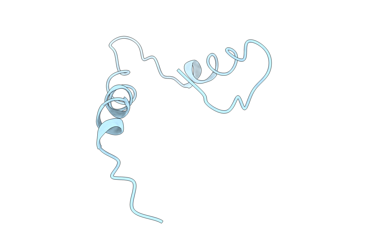
Solution Structure of the Human Immunodeficiency Virus Type 1 p6 Protein
Biological Source:
Source Organism:
Method Details:
Experimental Method:
Conformers Calculated:
104
Conformers Submitted:
1
Selection Criteria:
LEAST RESTRAINT VIOLATION
Macromolecular Entities
Structures with similar UniProt ID
Protein Blast
Polymer Type:polypeptide(L)
Molecule:PROTEIN P6
Gene (Uniprot):gag
Chain IDs:A
Chain Length:52
Number of Molecules:1
Biological Source:HUMAN IMMUNODEFICIENCY VIRUS TYPE 1
Ligand Molecules
Primary Citation
Solution Structure of the Human Immunodeficiency Virus Type 1 P6 Protein.
J.Biol.Chem. 280 42515 ? (2005)
PMID: 16234236 DOI: 10.1074/JBC.M507375200

Abstact

The human immunodeficiency virus type 1 p6 protein represents a docking site for several cellular and viral binding factors and fulfills major roles in the formation of infectious viruses. To date, however, the structure of this 52-amino acid protein, by far the smallest lentiviral protein known, either in its mature form as free p6 or as the C-terminal part of the Pr55 Gag polyprotein has not been unraveled. We have explored the high resolution structure and folding of p6 by CD and NMR spectroscopy. Under membranous solution conditions, p6 can adopt a helix-flexible helix structure; a short helix-1 (amino acids 14-18) is connected to a pronounced helix-2 (amino acids 33-44) by a flexible hinge region. Thus, p6 can be subdivided into two distinct structural and functional domains; helix-2 perfectly defines the region that binds to the virus budding factor AIP-1/ALIX, indicating that this structure is required for interaction with the endosomal sorting complex required for transport. The PTAP motif at the N terminus, comprising the primary late assembly domain, which is crucial for interaction with another cellular budding factor, Tsg101, does not exhibit secondary structure. However, the adjacent helix-1 may play an indirect role in the specific complex formation between p6 and the binding groove in Tsg101. Moreover, binding studies by NMR demonstrate that helix-2, which also comprises the LXXLF motif required for incorporation of the human immunodeficiency virus type 1 accessory protein Vpr into budding virions, specifically interacts with the Vpr binding region, indicating that under the specific solution conditions used for structure analysis, p6 adopted a functional conformation.

Legend

Protein

Chemical

Disease

Primary Citation of related structures