2C49 image
Deposition Date 2005-10-17
Release Date 2006-08-30
Last Version Date 2024-05-08
Entry Detail
PDB ID:
2C49
Keywords:
Title:
Crystal Structure of Methanocaldococcus jannaschii Nucleoside Kinase - An Archaeal Member of the Ribokinase Family
Biological Source:
Source Organism:
Host Organism:
Method Details:
Experimental Method:
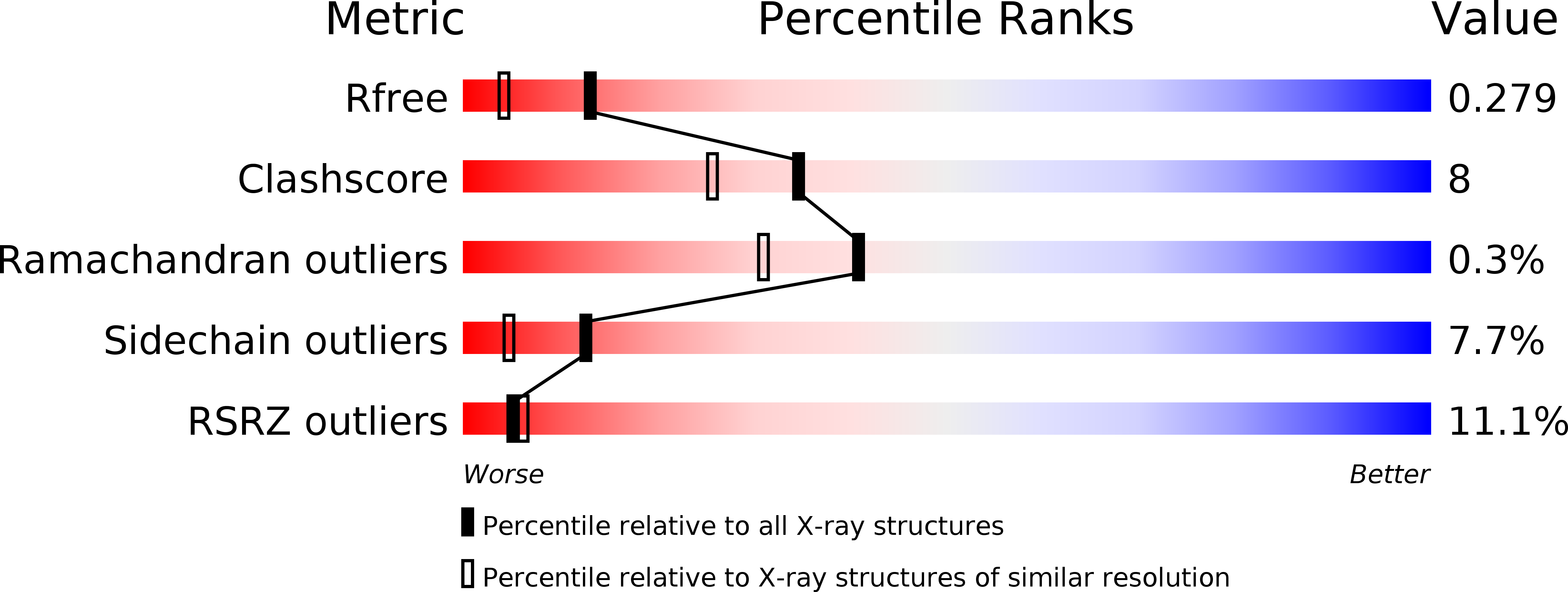
Resolution:
1.92 Å
R-Value Free:
0.28
R-Value Work:
0.24
R-Value Observed:
0.24
Space Group:
P 21 21 21
Macromolecular Entities
Structures with similar UniProt ID
Protein Blast
Polymer Type:polypeptide(L)
Molecule:SUGAR KINASE MJ0406
Gene (Uniprot):MJ0406
Chain IDs:A, B
Chain Length:302
Number of Molecules:2
Biological Source:METHANOCOCCUS JANNASCHII
Primary Citation
Structure of Methanocaldococcus Jannaschii Nucleoside Kinase: An Archaeal Member of the Ribokinase Family.
Acta Crystallogr.,Sect.D 62 1085 ? (2006)
PMID: 16929110 DOI: 10.1107/S0907444906024826

Abstact

Nucleoside kinase from the hyperthermophilic archaeon Methanocaldococcus jannaschii (MjNK) is a member of the ribokinase family. In the presence of ATP and Mg(2+), MjNK is able to catalyze the phosphorylation of a variety of nucleosides, including inosine, cytidine, guanosine and adenosine. Here, the crystal structure of MjNK, the first structure of an archaeal representative of the ribokinase family, is presented. The structure was solved using the multiple-wavelength anomalous dispersion technique. Three-dimensional structures of the unliganded enzyme and a complex of MjNK, an ATP analogue and adenosine were determined to 1.7 and 1.9 A resolution, respectively. Each subunit comprises an alpha/beta-domain and a smaller lid domain and has an overall fold characteristic of the ribokinase superfamily. MjNK shares highest structural similarity to the ribokinases from Escherichia coli and Thermotoga maritima. Similar to ribokinase and other superfamily members, the lid domain of MjNK undergoes a significant conformational change upon substrate binding. In the crystal structure of the MjNK complex, subunit A adopts a closed conformation and subunit B an open conformation. In subunit A all substrates and Mg(2+) were observed, whereas in subunit B only the ATP analogue could be clearly identified in the electron density. The structures of MjNK and E. coli ribokinase (EcRK) were compared with respect to putative determinants of thermal stability. Relative to EcRK, MjNK shows an increased charged and a decreased hydrophobic accessible surface area, as well as a higher fraction of charged residues, ionic networks and large aromatic clusters, characteristics that are frequently observed in enzymes from hyperthermophiles.

Legend

Protein

Chemical

Disease

Primary Citation of related structures