Deposition Date
2005-10-11
Release Date
2006-02-15
Last Version Date
2024-11-06
Entry Detail
PDB ID:
2C3M
Keywords:
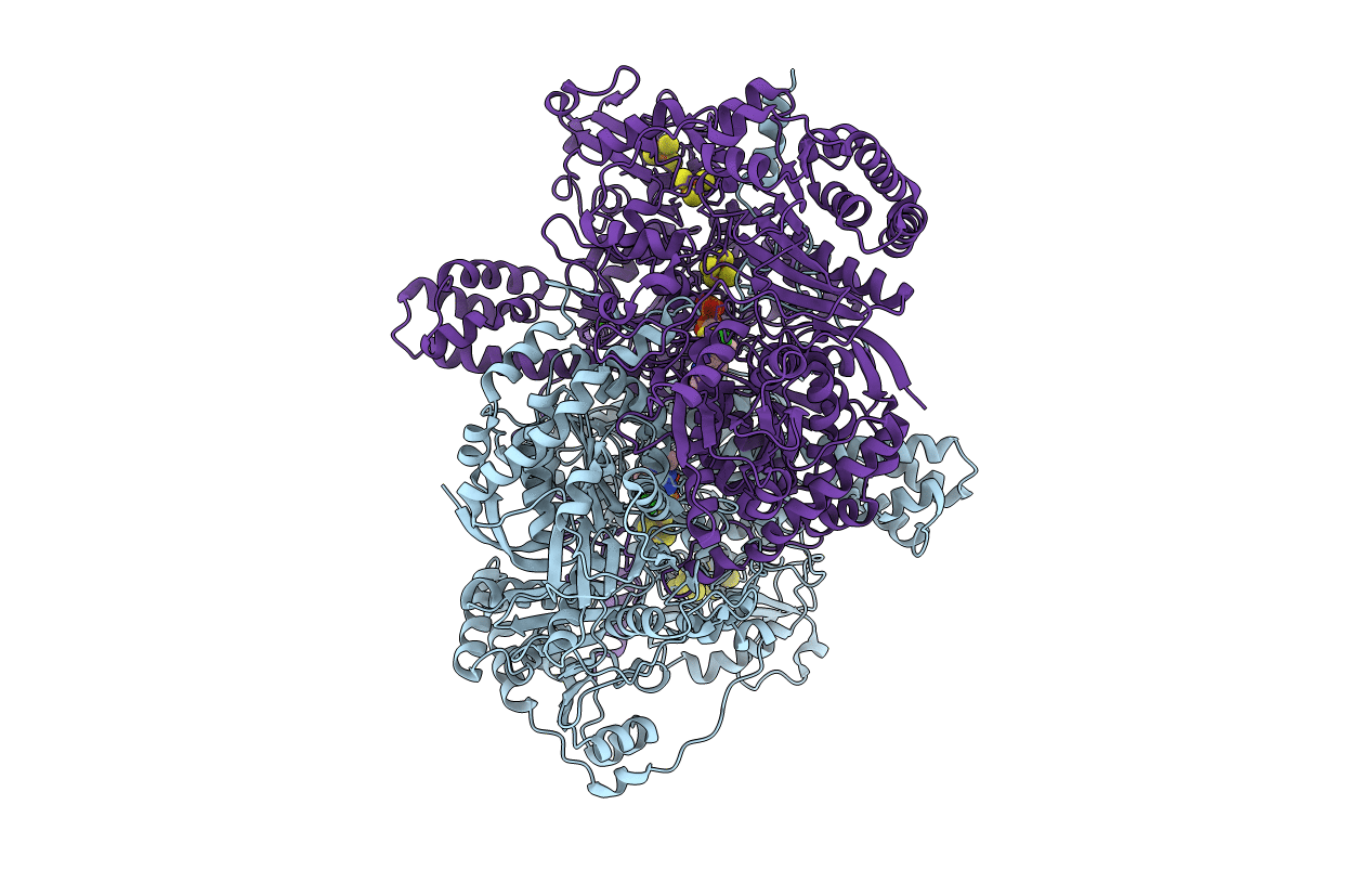
Title:
Crystal Structure Of Pyruvate-Ferredoxin Oxidoreductase From Desulfovibrio africanus
Biological Source:
Source Organism(s):
DESULFOVIBRIO AFRICANUS (Taxon ID: 873)
Method Details:
Experimental Method:
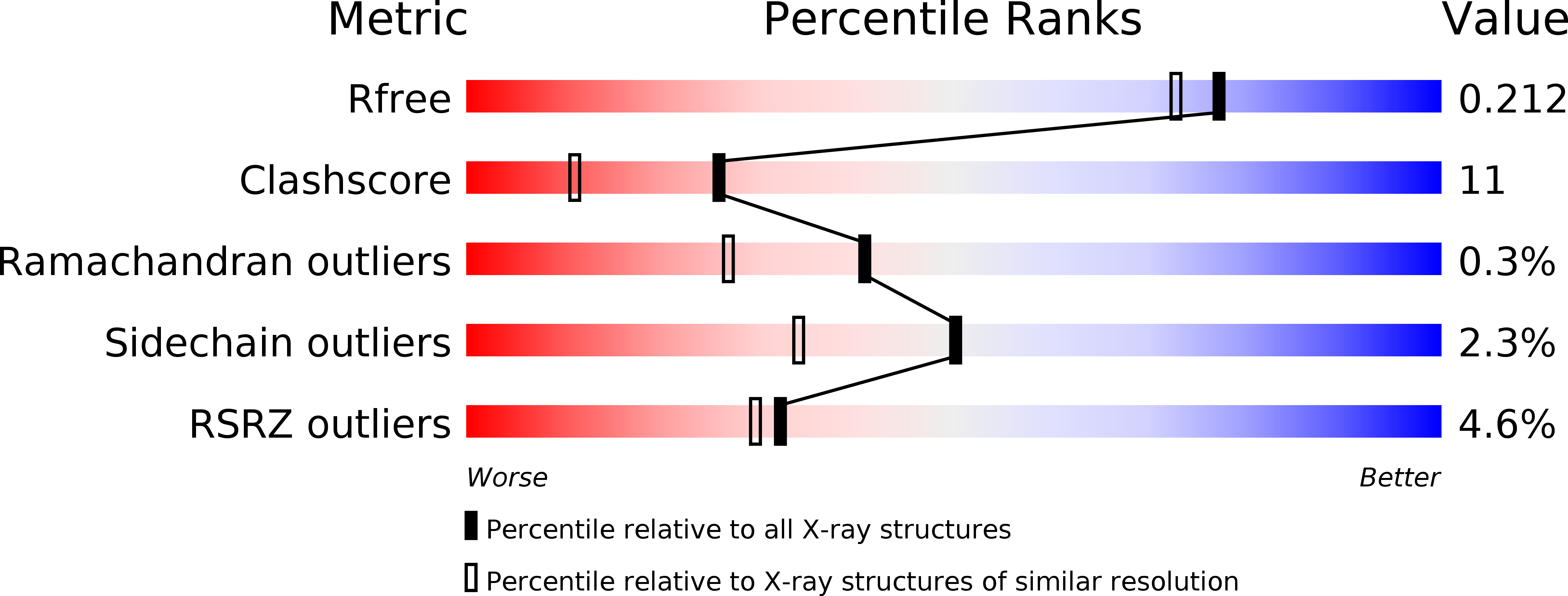
Resolution:
1.84 Å
R-Value Free:
0.22
R-Value Work:
0.19
R-Value Observed:
0.19
Space Group:
P 21 21 21