2C3B image
Deposition Date 2005-10-05
Release Date 2006-01-30
Last Version Date 2024-11-13
Entry Detail
PDB ID:
2C3B
Keywords:
Title:
The Crystal Structure of Aspergillus fumigatus Cyclophilin reveals 3D Domain Swapping of a Central Element
Biological Source:
Source Organism:
Host Organism:
Method Details:
Experimental Method:
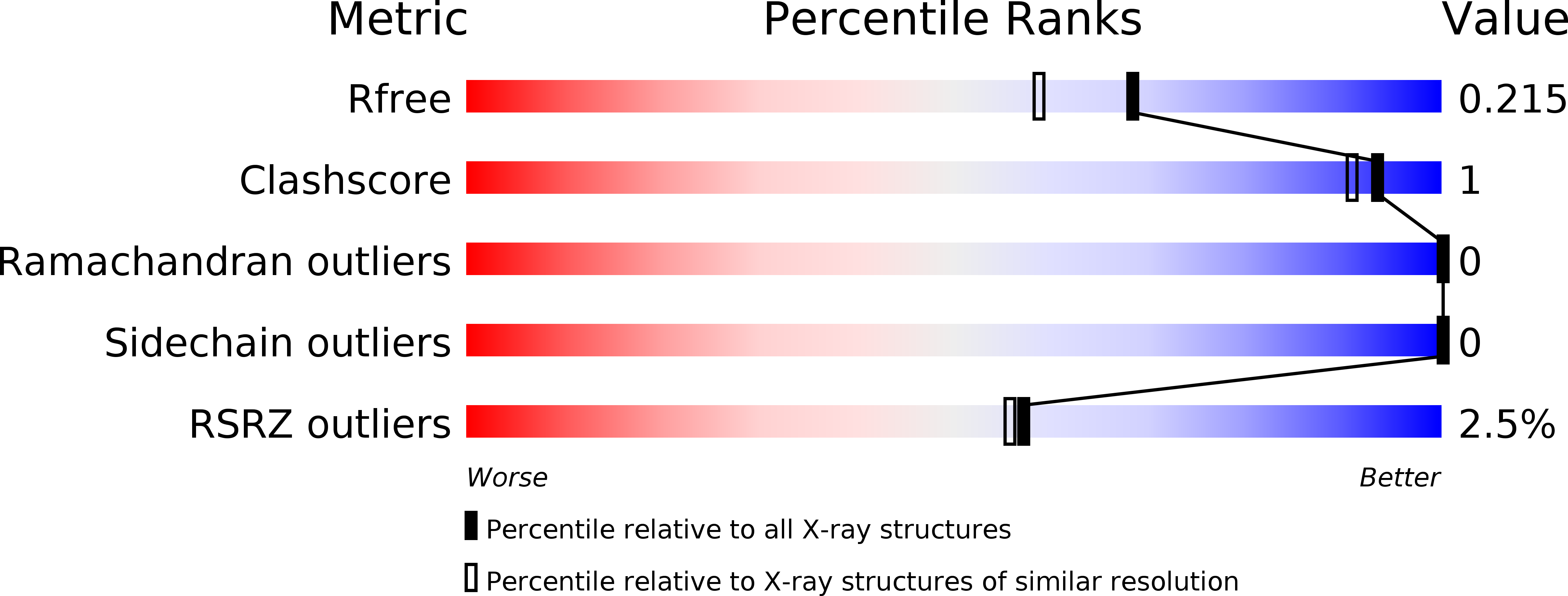
Resolution:
1.85 Å
R-Value Free:
0.21
R-Value Work:
0.18
R-Value Observed:
0.19
Space Group:
P 31 2 1
Macromolecular Entities
Polymer Type:polypeptide(L)
Molecule:PPIASE
Chain IDs:A, B
Chain Length:172
Number of Molecules:2
Biological Source:ASPERGILLUS FUMIGATUS
Ligand Molecules
Primary Citation
The Crystal Structure of Aspergillus Fumigatus Cyclophilin Reveals 3D Domain Swapping of a Central Element
Structure 14 185 ? (2006)
PMID: 16472738 DOI: 10.1016/J.STR.2005.10.015

Abstact

The crystal structure of Aspergillus fumigatus cyclophilin (Asp f 11) was solved by the multiwavelength anomalous dispersion method and was refined to a resolution of 1.85 A with R and R(free) values of 18.9% and 21.4%, respectively. Many cyclophilin structures have been solved to date, all showing the same monomeric conformation. In contrast, the structure of A. fumigatus cyclophilin reveals dimerization by 3D domain swapping and represents one of the first proteins with a swapped central domain. The domain-swapped element consists of two beta strands and a subsequent loop carrying a conserved tryptophan. The tryptophan binds into the active site, inactivating cis-trans isomerization. This might be a means of biological regulation. The two hinge loops leave the protein prone to misfolding. In this context, alternative forms of 3D domain swapping that can lead to N- or C-terminally swapped dimers, oligomers, and aggregates are discussed.

Legend

Protein

Chemical

Disease

Primary Citation of related structures