Deposition Date
2005-10-03
Release Date
2005-12-07
Last Version Date
2023-12-13
Entry Detail
PDB ID:
2C32
Keywords:
Title:
Co-axial association of recombinant eye lens aquaporin-0 observed in loosely packed 3D-crystals
Biological Source:
Source Organism(s):
BOS TAURUS (Taxon ID: 9913)
Expression System(s):
Method Details:
Experimental Method:
Resolution:
7.01 Å
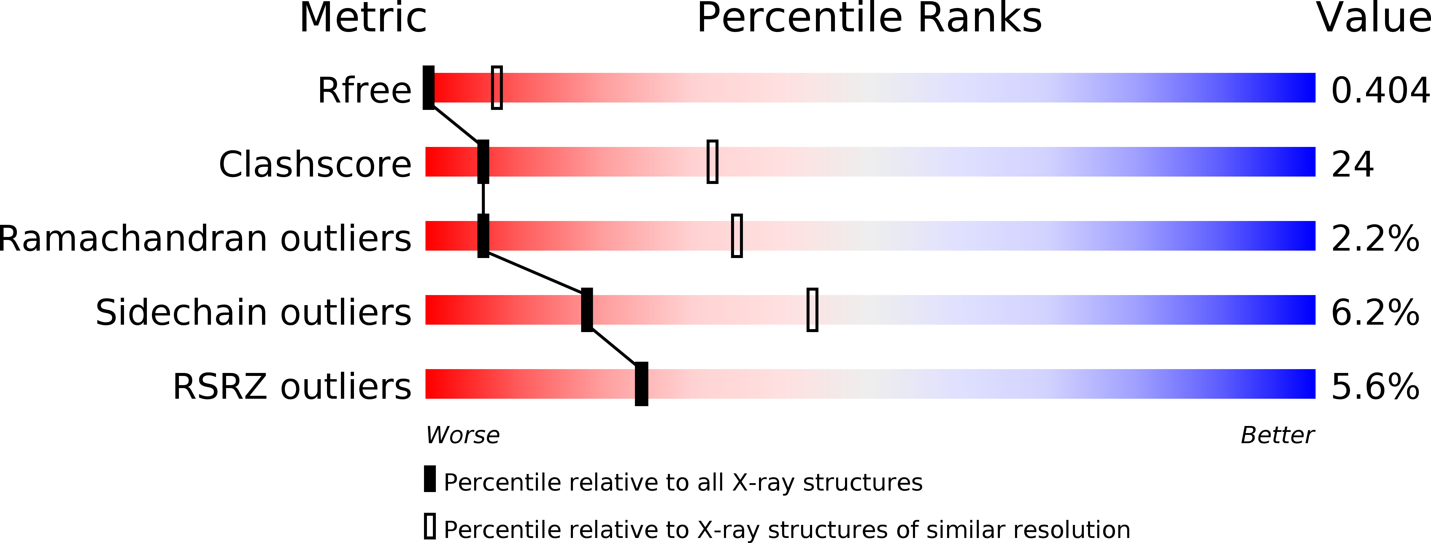
R-Value Free:
0.38
R-Value Work:
0.39
R-Value Observed:
0.39
Space Group:
I 4 3 2