2C29 image
Deposition Date 2005-09-27
Release Date 2006-10-16
Last Version Date 2024-05-08
Entry Detail
PDB ID:
2C29
Keywords:
Title:
Structure of dihydroflavonol reductase from Vitis vinifera at 1.8 A.
Biological Source:
Source Organism:
VITIS VINIFERA (Taxon ID: 29760)
Host Organism:
Method Details:
Experimental Method:
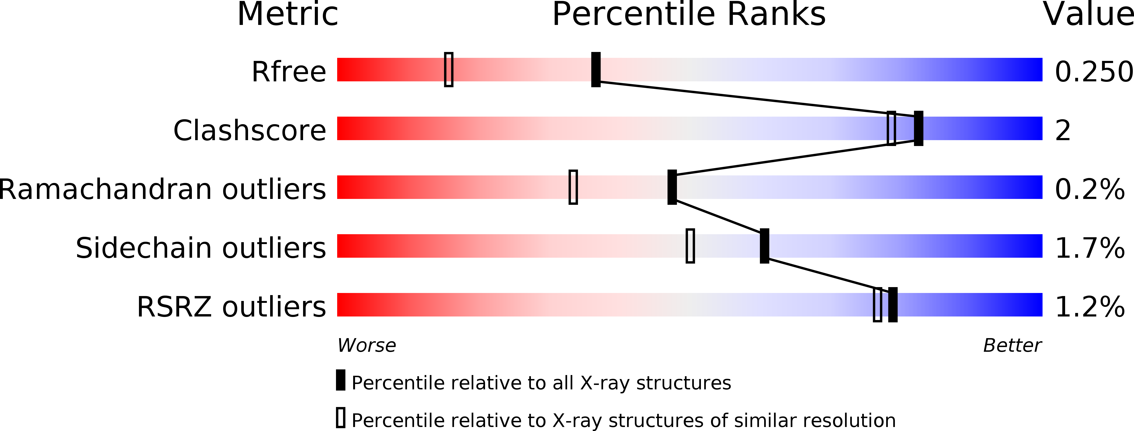
Resolution:
1.81 Å
R-Value Free:
0.24
R-Value Work:
0.19
R-Value Observed:
0.19
Space Group:
P 21 21 21
Macromolecular Entities
Structures with similar UniProt ID
Protein Blast
Polymer Type:polypeptide(L)
Molecule:DIHYDROFLAVONOL 4-REDUCTASE
Gene (Uniprot):dfr1
Chain IDs:A (auth: D), B (auth: F)
Chain Length:337
Number of Molecules:2
Biological Source:VITIS VINIFERA
Primary Citation
Crystal Structure of Grape Dihydroflavonol 4-Reductase, a Key Enzyme in Flavonoid Biosynthesis.
J.Mol.Biol. 368 1345 ? (2007)
PMID: 17395203 DOI: 10.1016/J.JMB.2007.02.088

Abstact

The nicotinamide adenine dinucleotide phosphate (NADPH)-dependent enzyme dihydroflavonol 4-reductase (DFR) catalyzes a late step in the biosynthesis of anthocyanins and condensed tannins, two flavonoid classes of importance to plant survival and human nutrition. This enzyme has been widely investigated in many plant species, but little is known about its structural and biochemical properties. To provide a basis for detailed structure-function studies, the crystal structure of Vitis vinifera DFR, heterologously expressed in Escherichia coli, has been determined at 1.8 A resolution. The 3D structure of the ternary complex obtained with the oxidized form of nicotinamide adenine dinucleotide phosphate and dihydroquercetin, one of the DFR substrates, presents common features with the short-chain dehydrogenase/reductase family, i.e., an N-terminal domain adopting a Rossmann fold and a variable C-terminal domain, which participates in substrate binding. The structure confirms the importance of the 131-156 region, which lines the substrate binding site and enlightens the role of a specific residue at position 133 (Asn or Asp), assumed to control substrate recognition. The activity of the wild-type enzyme and its variant N133D has been quantified in vitro, using dihydroquercetin or dihydrokaempferol. Our results demonstrate that position 133 cannot be solely responsible for the recognition of the B-ring hydroxylation pattern of dihydroflavonols.

Legend

Protein

Chemical

Disease

Primary Citation of related structures