Deposition Date
2005-09-21
Release Date
2006-01-13
Last Version Date
2024-10-16
Entry Detail
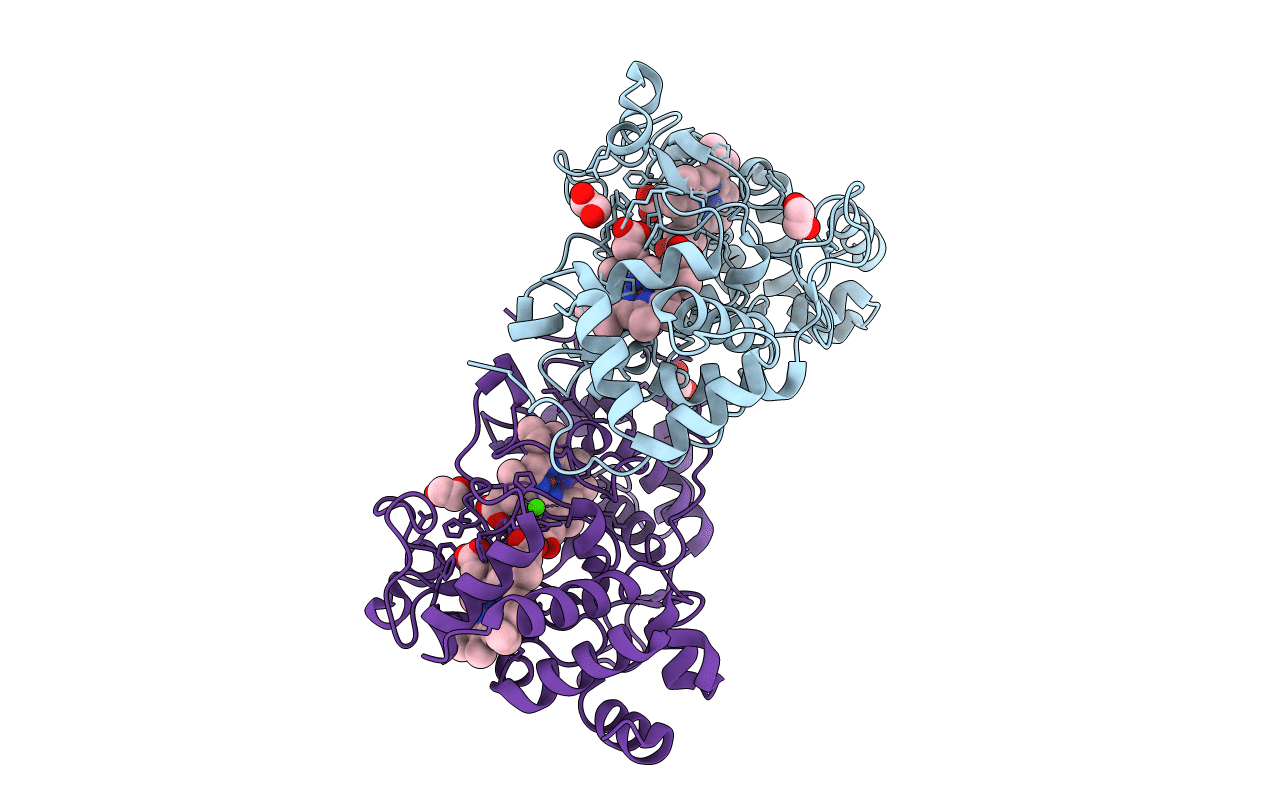
PDB ID:
2C1V
Keywords:
Title:
CRYSTAL STRUCTURE OF THE DI-HAEM CYTOCHROME C PEROXIDASE FROM PARACOCCUS PANTOTROPHUS - Mixed VALENCE FORM
Biological Source:
Source Organism(s):
PARACOCCUS PANTOTROPHUS (Taxon ID: 82367)
Method Details:
Experimental Method:
Resolution:
1.20 Å
R-Value Free:
0.18
R-Value Work:
0.17
R-Value Observed:
0.17
Space Group:
P 21 21 2