2BZR image
Deposition Date 2005-08-22
Release Date 2007-01-02
Last Version Date 2024-05-08
Entry Detail
PDB ID:
2BZR
Keywords:
Title:
Crystal structure of accD5 (Rv3280), an acyl-CoA carboxylase beta- subunit from Mycobacterium tuberculosis
Biological Source:
Source Organism:
Host Organism:
Method Details:
Experimental Method:
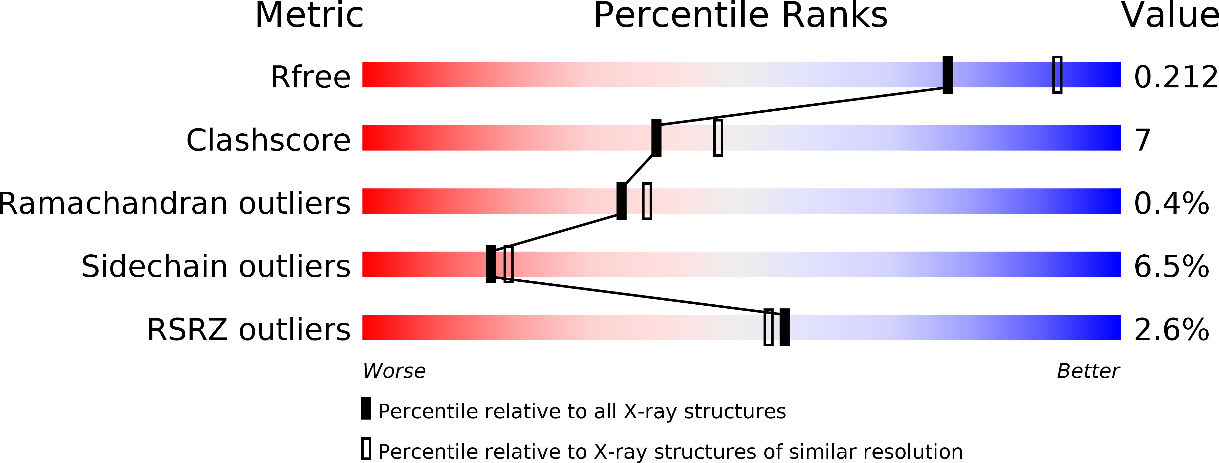
Resolution:
2.20 Å
R-Value Free:
0.21
R-Value Work:
0.17
R-Value Observed:
0.17
Space Group:
P 41 21 2
Macromolecular Entities
Polymer Type:polypeptide(L)
Molecule:PROPIONYL-COA CARBOXYLASE BETA CHAIN 5
Chain IDs:A, B, C, D, E, F
Chain Length:548
Number of Molecules:6
Biological Source:MYCOBACTERIUM TUBERCULOSIS
Primary Citation
Structural Diversity in the Six-Fold Redundant Set of Acyl-Coa Carboxyltransferases in Mycobacterium Tuberculosis.
FEBS Lett. 580 6898 ? (2006)
PMID: 17157300 DOI: 10.1016/J.FEBSLET.2006.11.054

Abstact

Mycobacterium tuberculosis contains multiple versions of the accA and accD genes that encode the alpha- and beta-subunits of at least three distinct multi-functional acyl-CoA carboxylase complexes. Because of its proposed involvement in pathogenic M. tuberculosis survival, the high-resolution crystal structure of the beta-subunit gene accD5 product has been determined and reveals a hexameric 356 kDa complex. Analysis of the active site properties of AccD5 and homology models of the other five M. tuberculosis AccD homologues reveals unexpected differences in their surface composition, providing a molecular rational key for a sorting mechanism governing correct acyl-CoA carboxylase holo complex assembly in M. tuberculosis.

Legend

Protein

Chemical

Disease

Primary Citation of related structures