Deposition Date
2005-08-03
Release Date
2005-10-05
Last Version Date
2023-12-13
Entry Detail
PDB ID:
2BYP
Keywords:
Title:
Crystal structure of Aplysia californica AChBP in complex with alpha- conotoxin ImI
Biological Source:
Source Organism(s):
APLYSIA CALIFORNICA (Taxon ID: 6500)
CONUS IMPERIALIS (Taxon ID: 35631)
CONUS IMPERIALIS (Taxon ID: 35631)
Expression System(s):
Method Details:
Experimental Method:
Resolution:
2.07 Å
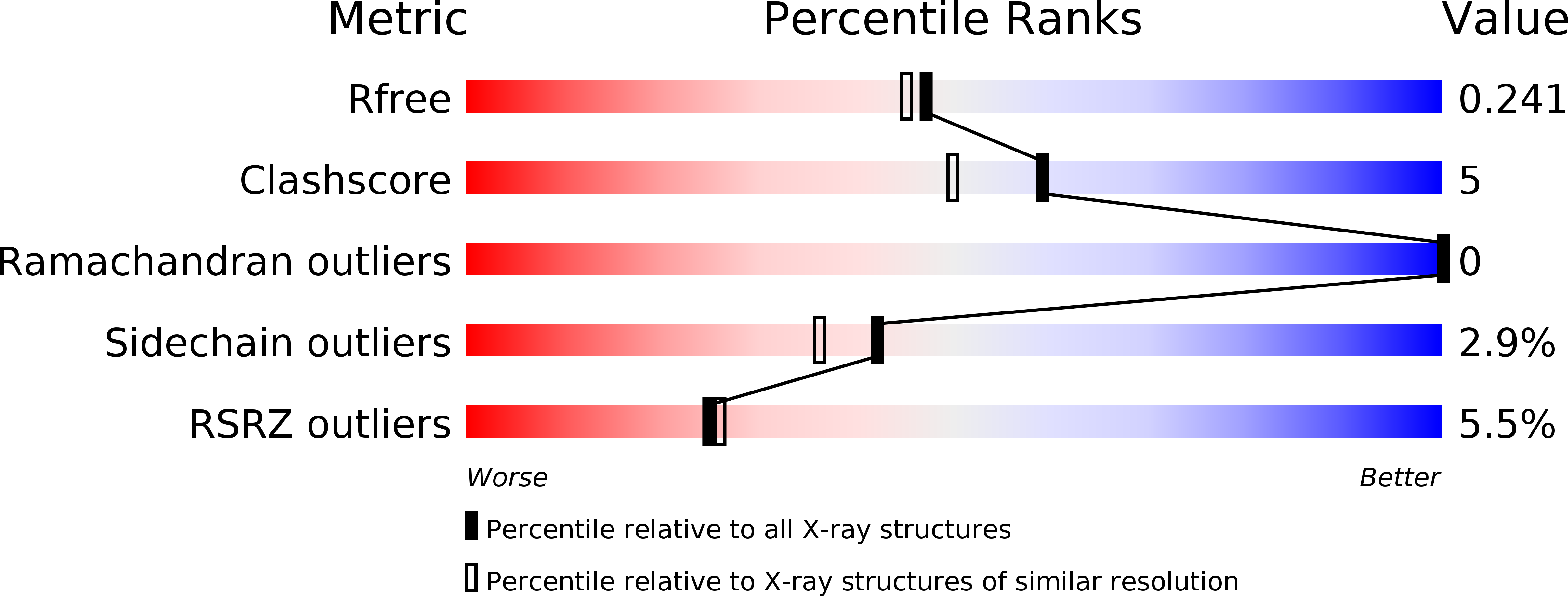
R-Value Free:
0.21
R-Value Work:
0.17
R-Value Observed:
0.17
Space Group:
I 2 2 2