2BX9 image
Deposition Date 2005-07-26
Release Date 2005-11-30
Last Version Date 2025-12-17
Entry Detail
PDB ID:
2BX9
Title:
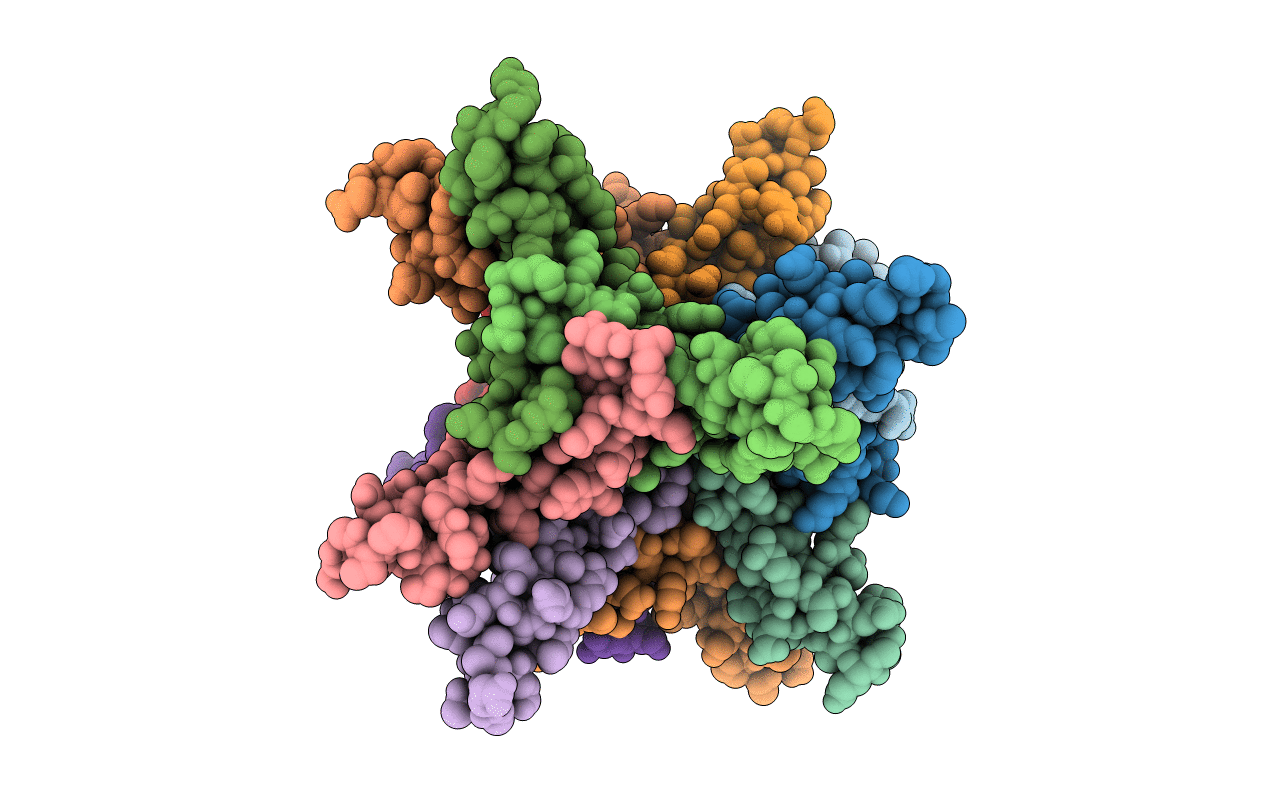
Crystal structure of B.subtilis Anti-TRAP protein, an antagonist of TRAP-RNA interactions
Biological Source:
Source Organism(s):
Expression System(s):
Method Details:
Experimental Method:
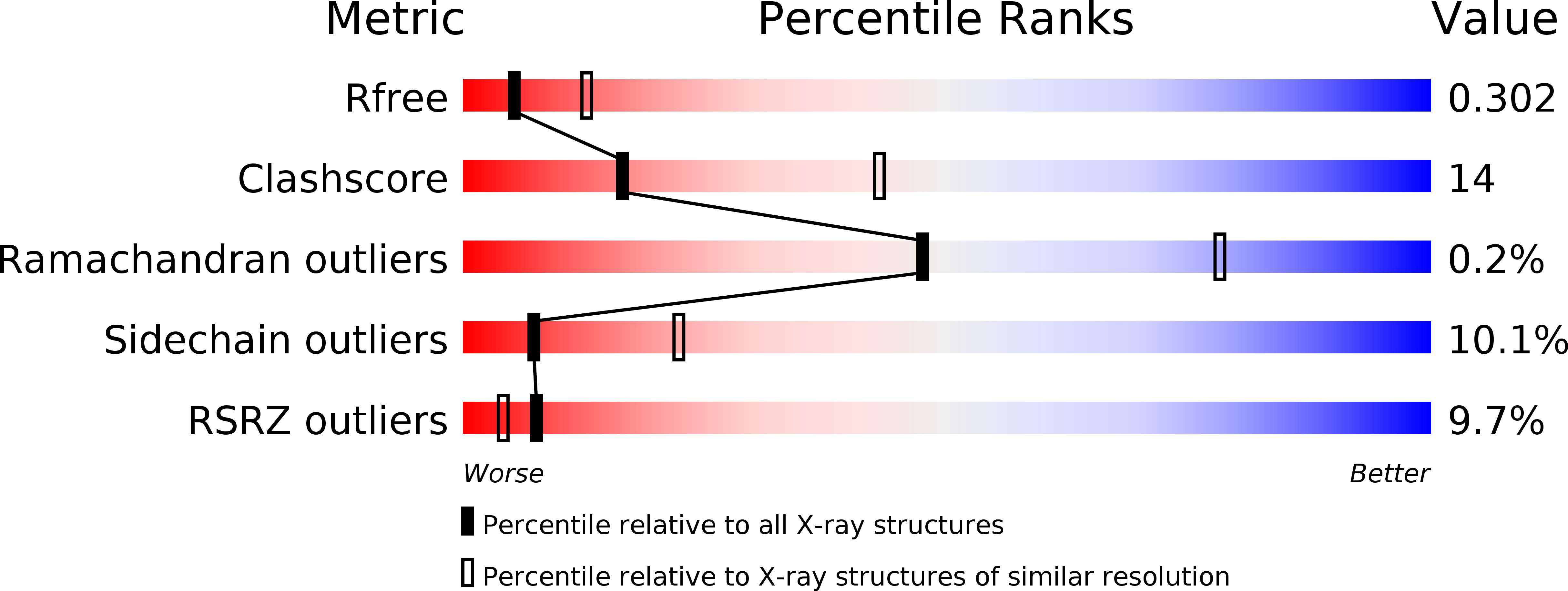
Resolution:
2.80 Å
R-Value Free:
0.29
R-Value Work:
0.19
R-Value Observed:
0.19
Space Group:
P 1
Macromolecular Entities
Structures with similar UniProt ID
Protein Blast
Polymer Type:polypeptide(L)
Molecule:TRYPTOPHAN RNA-BINDING ATTENUATOR PROTEIN-INHIBITORY PROTEIN
Gene (Uniprot):rtpA
Chain IDs:A, B, C, D, E, F, G, H, I, J, K, L
Chain Length:53
Number of Molecules:12
Biological Source:BACILLUS SUBTILIS
Ligand Molecules
Primary Citation
Crystal Structure of Bacillus Subtilis Anti-Trap Protein, an Antagonist of Trap/RNA Interaction.
Proc.Natl.Acad.Sci.USA 102 17600 ? (2005)
PMID: 16306262 DOI: 10.1073/PNAS.0508728102

Abstact

In Bacillus subtilis the anti-TRAP protein (AT) is produced in response to the accumulation of uncharged tRNA(Trp). AT regulates expression of genes involved in tryptophan biosynthesis and transport by binding to the tryptophan-activated trp RNA-binding attenuation protein (TRAP) and preventing its interaction with several mRNAs. Here, we report the x-ray structure of AT at 2.8 angstroms resolution, showing that the protein subunits assemble into tight trimers. Four such trimers are further associated into a 12-subunit particle in which individual trimers are related by twofold and threefold symmetry axes. Twelve DnaJ-like, cysteine-rich zinc-binding domains form spikes on the surface of the dodecamer. Available data suggest several possible ways for AT to interact with the 11-subunit TRAP. Interaction between the two symmetry-mismatching molecules could be assisted by the flexible nature of AT zinc-binding domains.

Legend

Protein

Chemical

Disease

Primary Citation of related structures
Feedback Form
Name
Email
Institute
Feedback