2BW0 image
Deposition Date 2005-07-07
Release Date 2005-07-08
Last Version Date 2023-12-13
Entry Detail
PDB ID:
2BW0
Keywords:
Title:
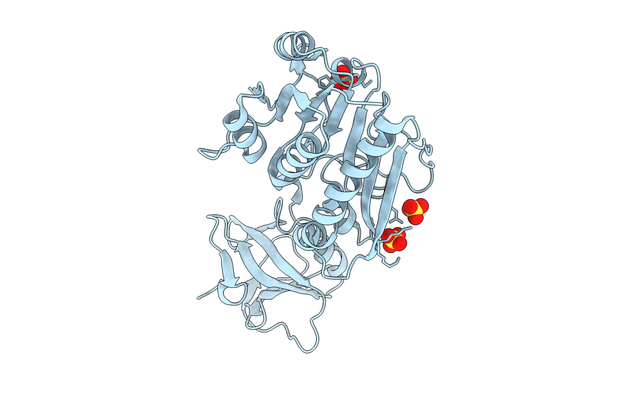
Crystal Structure of the hydrolase domain of Human 10-Formyltetrahydrofolate 2 dehydrogenase
Biological Source:
Source Organism:
HOMO SAPIENS (Taxon ID: 9606)
Host Organism:
Method Details:
Experimental Method:
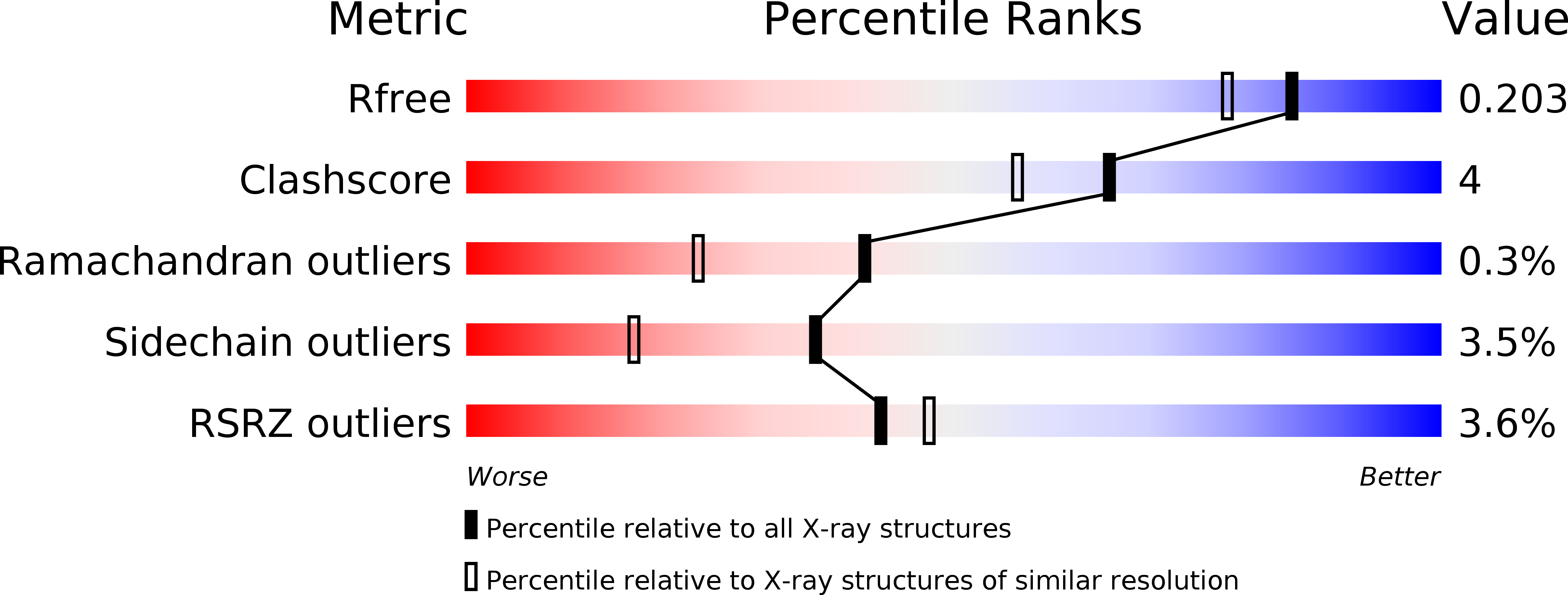
Resolution:
1.70 Å
R-Value Free:
0.20
R-Value Work:
0.17
R-Value Observed:
0.17
Space Group:
C 1 2 1
Macromolecular Entities
Structures with similar UniProt ID
Protein Blast
Polymer Type:polypeptide(L)
Molecule:10-FORMYLTETRAHYDROFOLATE DEHYDROGENASE
Gene (Uniprot):ALDH1L1
Chain IDs:A
Chain Length:329
Number of Molecules:1
Biological Source:HOMO SAPIENS
Ligand Molecules
Primary Citation
Structures of the Hydrolase Domain of Human 10-Formyltetrahydrofolate Dehydrogenase and its Complex with a Substrate Analogue.
Acta Crystallogr.,Sect.D 62 1294 ? (2006)
PMID: 17057331 DOI: 10.1107/S0907444906026849

Abstact

10-Formyltetrahydrofolate dehydrogenase is a ubiquitously expressed enzyme in the human body. It catalyses the formation of tetrahydrofolate and carbon dioxide from 10-formyltetrahydrofolate, thereby playing an important role in the human metabolism of one-carbon units. It is a two-domain protein in which the N-terminal domain hydrolyses 10-formyltetrahydrofolate into formate and tetrahydrofolate. The high-resolution crystal structure of the hydrolase domain from human 10-formyltetrahydrofolate dehydrogenase has been determined in the presence and absence of a substrate analogue. The structures reveal conformational changes of two loops upon ligand binding, while key active-site residues appear to be pre-organized for catalysis prior to substrate binding. Two water molecules in the structures mark the positions of key oxygen moieties in the catalytic reaction and reaction geometries are proposed based on the structural data.

Legend

Protein

Chemical

Disease

Primary Citation of related structures