2BVG image
Entry Detail
PDB ID:
2BVG
Keywords:
Title:
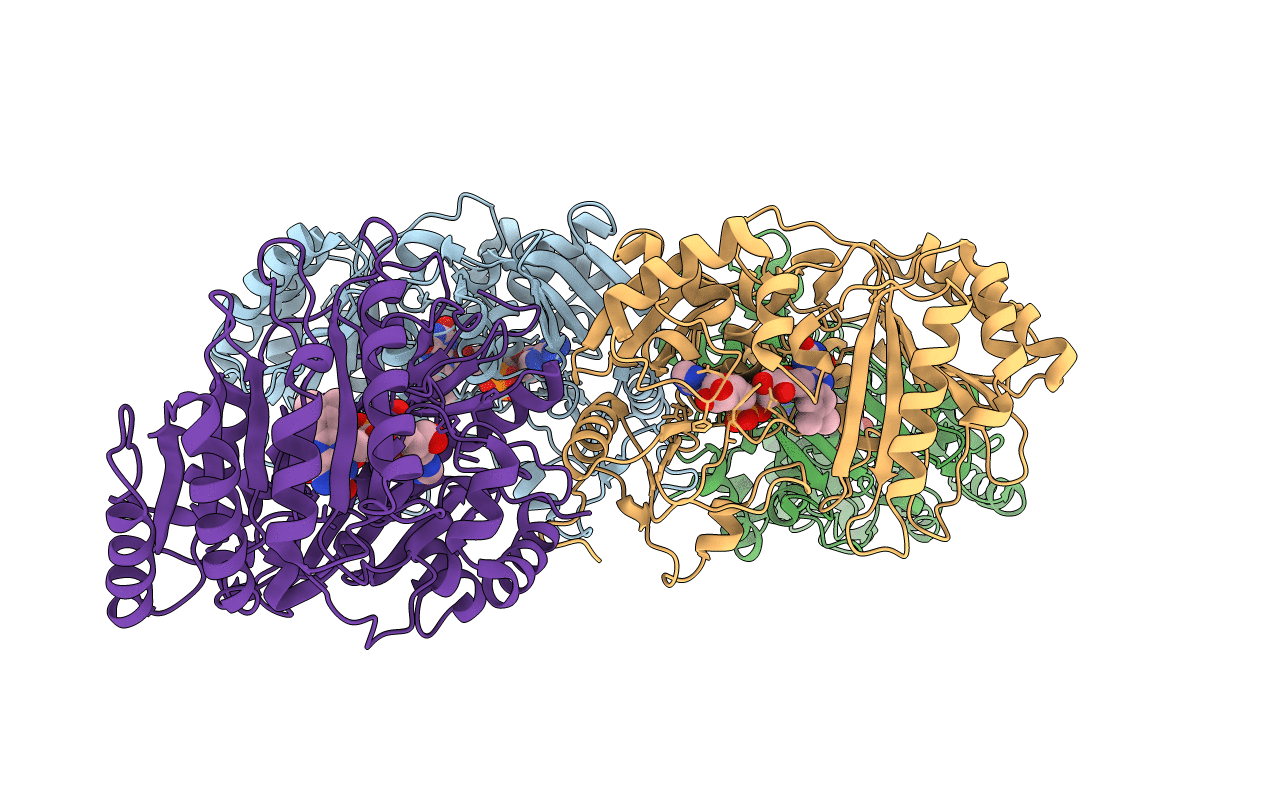
Crystal structure of 6-hydoxy-D-nicotine oxidase from Arthrobacter nicotinovorans. Crystal Form 1 (P21)
Biological Source:
Host Organism:
PDB Version:
Deposition Date:
2005-06-28
Release Date:
2005-08-17
Method Details:
Experimental Method:
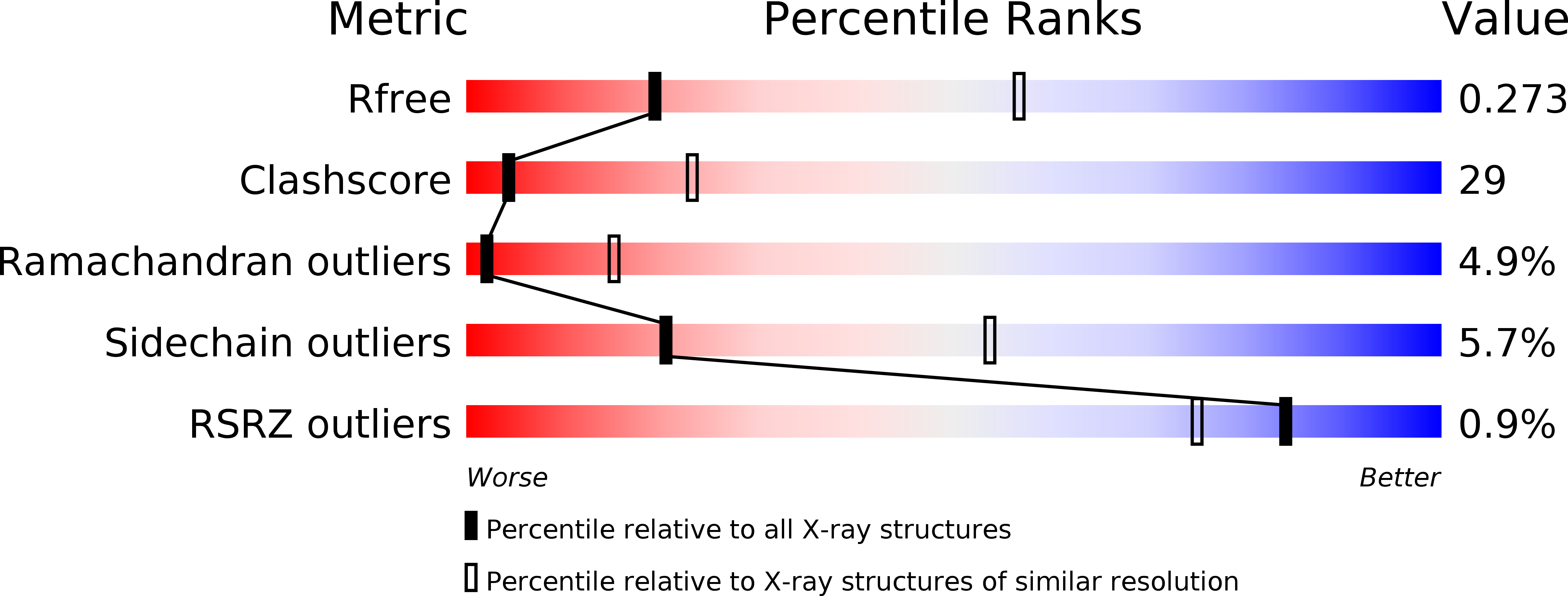
Resolution:
3.18 Å
R-Value Free:
0.28
R-Value Work:
0.25
R-Value Observed:
0.25
Space Group:
P 1 21 1
Macromolecular Entities
Polymer Type:polypeptide(L)
Description:6-HYDROXY-D-NICOTINE OXIDASE
Mutations:YES
Chain IDs:A, B, C, D
Chain Length:459
Number of Molecules:4
Biological Source:ARTHROBACTER NICOTINOVORANS
Ligand Molecules
Primary Citation
Crystal Structure of 6-Hydroxy-D-Nicotine Oxidase from Arthrobacter Nicotinovorans.
J.Mol.Biol. 352 418 ? (2005)
PMID: 16095622 DOI: 10.1016/J.JMB.2005.07.041

Abstact

The crystal structure of 6-hydroxy-d-nicotine oxidase (EC 1.5.3.6) was solved by X-ray diffraction analysis in three crystal forms at resolutions up to 1.9 A. The enzyme is monomeric in solution and also in the mother liquor but formed disulfide-dimers in all crystals. It belongs to the p-cresol methylhydroxylase-vanillyl-alcohol oxidase family and contains an FAD covalently bound to the polypeptide. The covalent bond of this enzyme was the first for which a purely autocatalytic formation had been shown. In contrast to previous reports, the bond does not involve N(epsilon2) (N3) of His72 but the N(delta1) (N1) atom. The geometry of this reaction is proposed and the autoflavinylation is discussed in the light of homologous structures. The enzyme is specific for 6-hydroxy-D-nicotine and is inhibited by the L-enantiomer. This observation was verified by modeling enzyme-substrate and enzyme-inhibitor complexes, which also showed the geometry of the catalyzed reaction. The binding models indicated that the deprotonation of the substrate rather than the hydride transfer is the specificity-determining step. The functionally closely related 6-hydroxy-L-nicotine oxidase processing the L-enantiomer is sequence-related to the greater glutathione reductase family with quite a different chainfold. A model of this "sister enzyme" derived from known homologous structures suggests that the reported L-substrate specificity and D-enantiomer inhibition are also determined by the location of the deprotonating base.

Legend

Protein

Chemical

Disease

Primary Citation of related structures