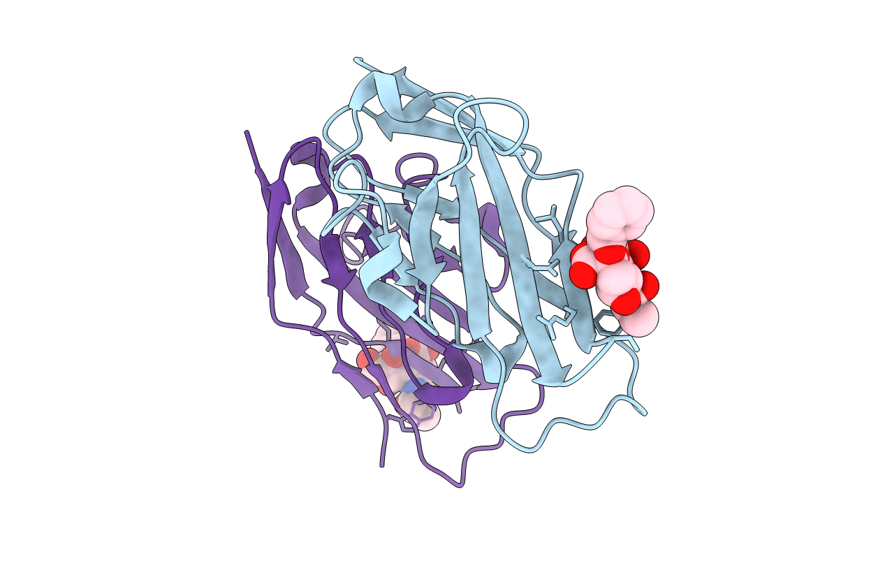
2BVE image
Entry Detail
PDB ID:
2BVE
Keywords:
Title:
Structure of the N-terminal of Sialoadhesin in complex with 2-Phenyl- Prop5Ac
Biological Source:
Source Organism:
Host Organism:
PDB Version:
Deposition Date:
2005-06-27
Release Date:
2006-07-19
Method Details:
Experimental Method:
Resolution:
2.20 Å
R-Value Free:
0.27
R-Value Work:
0.20
R-Value Observed:
0.20
Space Group:
P 21 21 21
Macromolecular Entities
Polymer Type:polypeptide(L)
Description:SIALOADHESIN
Chain IDs:A, B
Chain Length:119
Number of Molecules:2
Biological Source:MUS MUSCULUS
Ligand Molecules
Primary Citation
Crystallographic and in Silico Analysis of the Sialoside-Binding Characteristics of the Siglec Sialoadhesin.
J.Mol.Biol. 365 1469 ? (2007)
PMID: 17137591 DOI: 10.1016/J.JMB.2006.10.084

Abstact

The Siglec family of receptors mediates cell-surface interactions through recognition of sialylated glycoconjugates. Previously reported structures of the N-terminal domain of the Siglec sialoadhesin (SnD1) in complex with various sialic acid analogs revealed the structural template for sialic acid binding. To characterize further the carbohydrate-binding properties, we have determined the crystal structures of SnD1 in the absence of ligand, and in complex with 2-benzyl-Neu5NPro and 2-benzyl-Neu5NAc. These structures reveal that SnD1 undergoes very few structural changes on ligand binding and detail how two novel classes of sialic acid analogs bind, one of which unexpectedly can induce Siglec dimerization. In conjunction with in silico analysis, this set of structures informs us about the design of putative ligands with enhanced binding affinities and specificities to different Siglecs, and provides data with which to test the effectiveness of different computational drug design protocols.

Legend

Protein

Chemical

Disease

Primary Citation of related structures