2BV5 image
Deposition Date 2005-06-22
Release Date 2005-07-14
Last Version Date 2024-10-16
Entry Detail
PDB ID:
2BV5
Keywords:
Title:
CRYSTAL STRUCTURE OF THE HUMAN PROTEIN TYROSINE PHOSPHATASE PTPN5 AT 1.8A RESOLUTION
Biological Source:
Source Organism:
HOMO SAPIENS (Taxon ID: 9606)
Method Details:
Experimental Method:
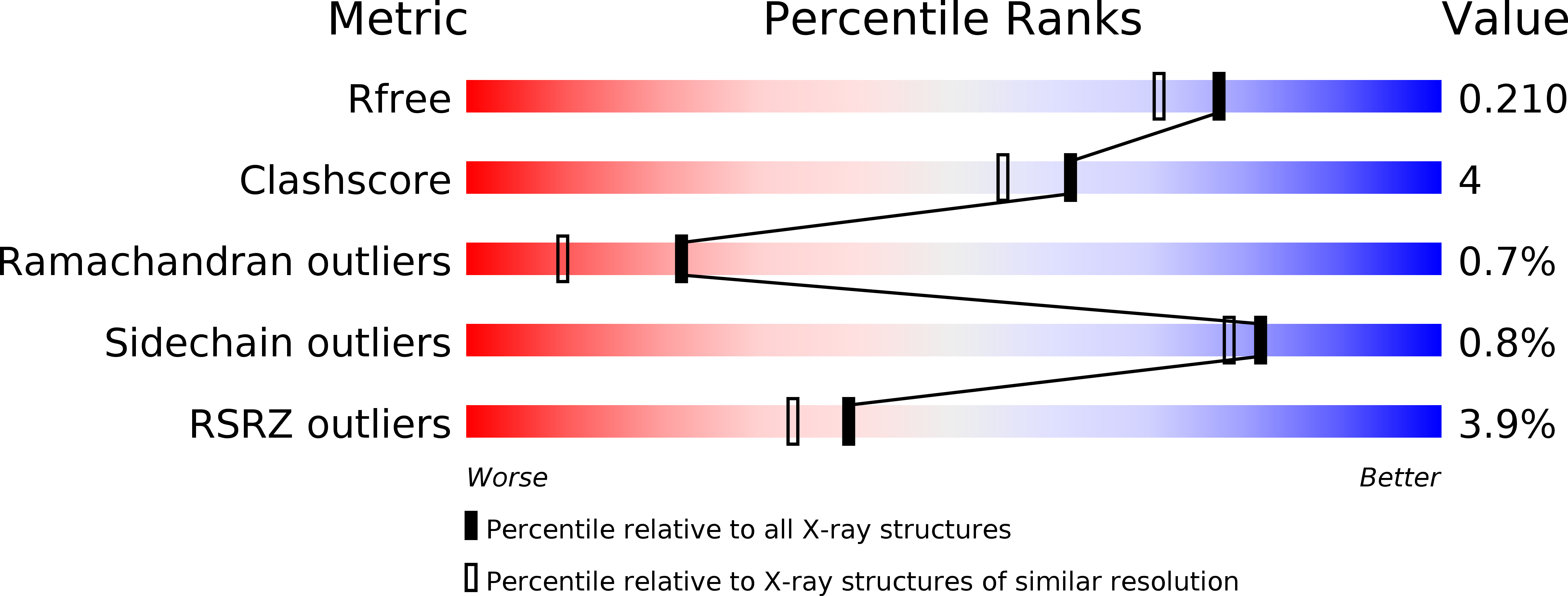
Resolution:
1.80 Å
R-Value Free:
0.20
R-Value Work:
0.16
R-Value Observed:
0.16
Space Group:
P 21 21 21
Macromolecular Entities
Polymer Type:polypeptide(L)
Molecule:TYROSINE-PROTEIN PHOSPHATASE, NON-RECEPTOR TYPE 5
Gene (Uniprot):PTPN5
Mutations:YES
Chain IDs:A
Chain Length:282
Number of Molecules:1
Biological Source:HOMO SAPIENS
Modified Residue
Compound ID Chain ID Parent Comp ID Details 2D Image
SCY A CYS S-ACETYL-CYSTEINE
Primary Citation
Crystal structures and inhibitor identification for PTPN5, PTPRR and PTPN7: a family of human MAPK-specific protein tyrosine phosphatases.
Biochem. J. 395 483 491 (2006)
PMID: 16441242 DOI: 10.1042/BJ20051931

Abstact

Protein tyrosine phosphatases PTPN5, PTPRR and PTPN7 comprise a family of phosphatases that specifically inactivate MAPKs (mitogen-activated protein kinases). We have determined high-resolution structures of all of the human family members, screened them against a library of 24000 compounds and identified two classes of inhibitors, cyclopenta[c]quinolinecarboxylic acids and 2,5-dimethylpyrrolyl benzoic acids. Comparative structural analysis revealed significant differences within this conserved family that could be explored for the design of selective inhibitors. PTPN5 crystallized, in two distinct crystal forms, with a sulphate ion in close proximity to the active site and the WPD (Trp-Pro-Asp) loop in a unique conformation, not seen in other PTPs, ending in a 3(10)-helix. In the PTPN7 structure, the WPD loop was in the closed conformation and part of the KIM (kinase-interaction motif) was visible, which forms an N-terminal aliphatic helix with the phosphorylation site Thr66 in an accessible position. The WPD loop of PTPRR was open; however, in contrast with the structure of its mouse homologue, PTPSL, a salt bridge between the conserved lysine and aspartate residues, which has been postulated to confer a more rigid loop structure, thereby modulating activity in PTPSL, does not form in PTPRR. One of the identified inhibitor scaffolds, cyclopenta[c]quinoline, was docked successfully into PTPRR, suggesting several possibilities for hit expansion. The determined structures together with the established SAR (structure-activity relationship) propose new avenues for the development of selective inhibitors that may have therapeutic potential for treating neurodegenerative diseases in the case of PTPRR or acute myeloblastic leukaemia targeting PTPN7.

Legend

Protein

Chemical

Disease

Primary Citation of related structures