Deposition Date
2005-06-17
Release Date
2006-09-05
Last Version Date
2023-12-13
Entry Detail
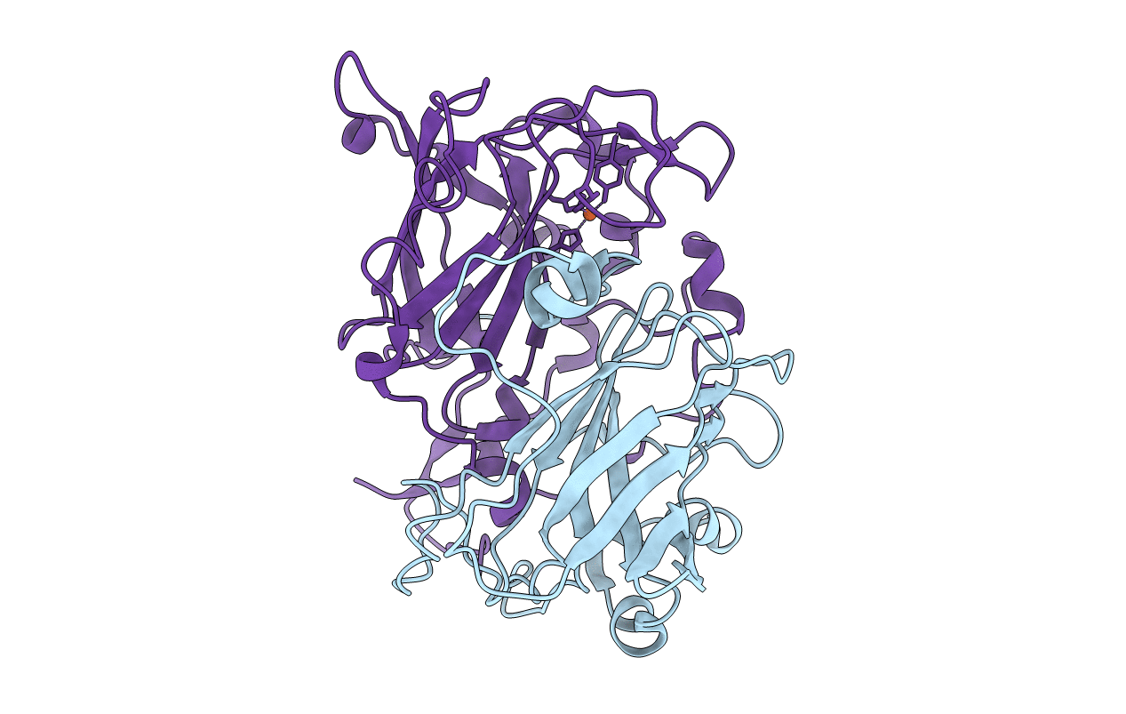
PDB ID:
2BUT
Keywords:
Title:
Crystal Structure Of Protocatechuate 3,4-Dioxygenase from Acinetobacter Sp. ADP1 Mutant R457S - APO
Biological Source:
Source Organism(s):
ACINETOBACTER CALCOACETICUS (Taxon ID: 62977)
Expression System(s):
Method Details:
Experimental Method:
Resolution:
1.85 Å
R-Value Free:
0.21
R-Value Work:
0.17
R-Value Observed:
0.17
Space Group:
I 2 3