2BTQ image
Deposition Date 2005-06-06
Release Date 2005-06-14
Last Version Date 2023-12-13
Entry Detail
PDB ID:
2BTQ
Title:
Structure of BtubAB heterodimer from Prosthecobacter dejongeii
Biological Source:
Source Organism:
Method Details:
Experimental Method:
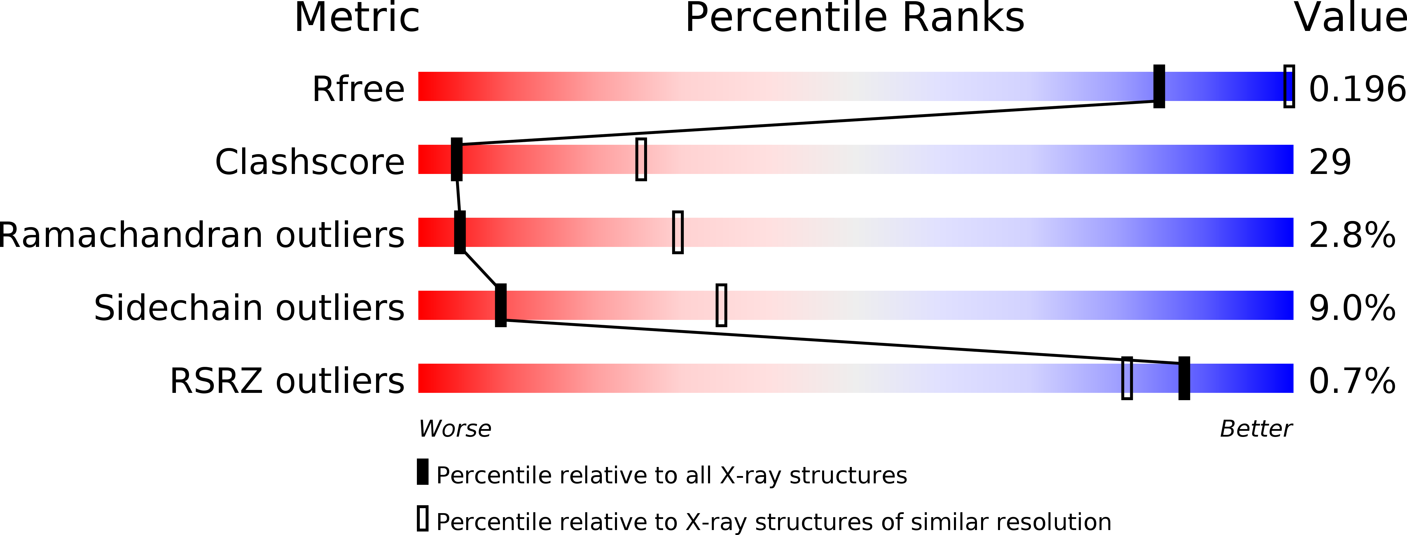
Resolution:
3.20 Å
R-Value Free:
0.24
R-Value Work:
0.21
R-Value Observed:
0.21
Space Group:
P 65 2 2
Macromolecular Entities
Structures with similar UniProt ID
Protein Blast
Polymer Type:polypeptide(L)
Molecule:TUBULIN BTUBA
Gene (Uniprot):btubA
Chain IDs:A
Chain Length:473
Number of Molecules:1
Biological Source:PROSTHECOBACTER DEJONGEII
Structures with similar UniProt ID
Protein Blast
Polymer Type:polypeptide(L)
Molecule:TUBULIN BTUBB
Gene (Uniprot):btubB
Chain IDs:B
Chain Length:426
Number of Molecules:1
Biological Source:PROSTHECOBACTER DEJONGEII
Primary Citation
Structure of Bacterial Tubulin Btuba/B: Evidence for Horizontal Gene Transfer.
Proc.Natl.Acad.Sci.USA 102 9170 ? (2005)
PMID: 15967998 DOI: 10.1073/PNAS.0502859102

Abstact

alphabeta-Tubulin heterodimers, from which the microtubules of the cytoskeleton are built, have a complex chaperone-dependent folding pathway. They are thought to be unique to eukaryotes, whereas the homologue FtsZ can be found in bacteria. The exceptions are BtubA and BtubB from Prosthecobacter, which have higher sequence homology to eukaryotic tubulin than to FtsZ. Here we show that some of their properties are different from tubulin, such as weak dimerization and chaperone-independent folding. However, their structure is strikingly similar to tubulin including surface loops, and BtubA/B form tubulin-like protofilaments. Presumably, BtubA/B were transferred from a eukaryotic cell by horizontal gene transfer because their high degree of similarity to eukaryotic genes is unique within the Prosthecobacter genome.

Legend

Protein

Chemical

Disease

Primary Citation of related structures
Feedback Form
Name
Email
Institute
Feedback