Deposition Date
2005-05-14
Release Date
2005-12-13
Last Version Date
2024-11-20
Entry Detail
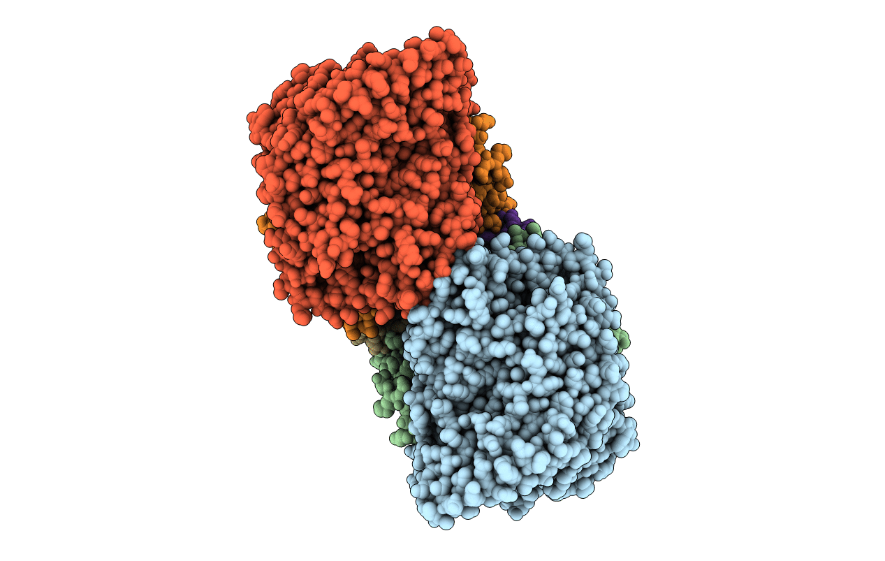
PDB ID:
2BS4
Keywords:
Title:
GLU C180 -> ILE VARIANT QUINOL:FUMARATE REDUCTASE FROMWOLINELLA SUCCINOGENES
Biological Source:
Source Organism(s):
WOLINELLA SUCCINOGENES (Taxon ID: 844)
Method Details:
Experimental Method:
Resolution:
2.76 Å
R-Value Free:
0.21
R-Value Work:
0.20
R-Value Observed:
0.20
Space Group:
P 1 21 1