
Deposition Date
2005-05-13
Release Date
2005-10-26
Last Version Date
2024-05-08
Entry Detail
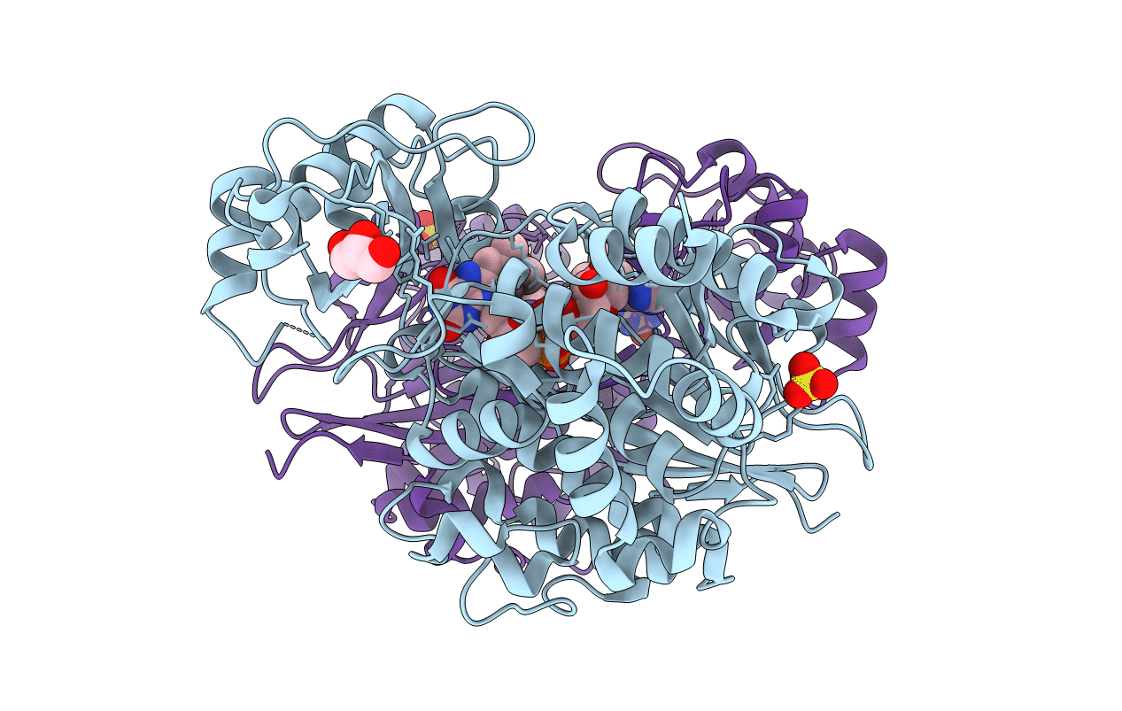
PDB ID:
2BRY
Keywords:
Title:
Crystal structure of the native monooxygenase domain of MICAL at 1.45 A resolution
Biological Source:
Source Organism(s):
MUS MUSCULUS (Taxon ID: 10090)
Expression System(s):
Method Details:
Experimental Method:
Resolution:
1.45 Å
R-Value Free:
0.22
R-Value Work:
0.18
R-Value Observed:
0.18
Space Group:
P 1 21 1
