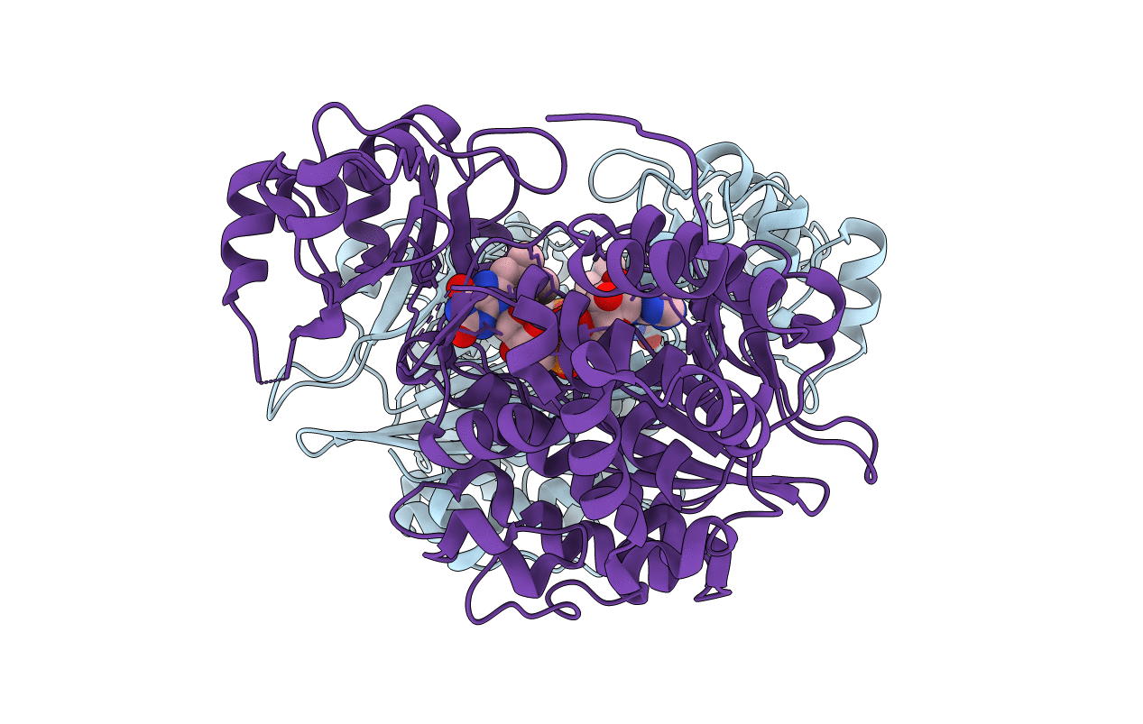
2BRA image
Deposition Date 2005-05-04
Release Date 2005-11-01
Last Version Date 2024-05-08
Entry Detail
PDB ID:
2BRA
Keywords:
Title:
Structure of N-Terminal FAD Binding motif of mouse MICAL
Biological Source:
Source Organism:
MUS MUSCULUS (Taxon ID: 10090)
Method Details:
Experimental Method:
Resolution:
2.00 Å
R-Value Free:
0.26
R-Value Work:
0.19
R-Value Observed:
0.19
Space Group:
P 1 21 1
Macromolecular Entities
Structures with similar UniProt ID
Protein Blast
Polymer Type:polypeptide(L)
Molecule:NEDD9 INTERACTING PROTEIN WITH CALPONIN HOMOLOGY AND LIM DOMAINS
Gene (Uniprot):Mical1
Mutagens:YES
Chain IDs:A, B
Chain Length:484
Number of Molecules:2
Biological Source:MUS MUSCULUS
Primary Citation
Structure and activity of the axon guidance protein MICAL.
Proc. Natl. Acad. Sci. U.S.A. 102 16830 16835 (2005)
PMID: 16275926 DOI: 10.1073/pnas.0504838102

Abstact

During development, neurons are guided to their targets by short- and long-range attractive and repulsive cues. MICAL, a large multidomain protein, is required for the combined action of semaphorins and plexins in axon guidance. Here, we present the structure of the N-terminal region of MICAL (MICAL(fd)) determined by x-ray diffraction to 2.0 A resolution. The structure shows that MICAL(fd) is an FAD-containing module structurally similar to aromatic hydroxylases and amine oxidases. In addition, we present biochemical data that show that MICAL(fd) is a flavoenzyme that in the presence of NADPH reduces molecular oxygen to H(2)O(2) (K(m,NAPDH) = 222 microM; k(cat) = 77 sec(-1)), a molecule with known signaling properties. We propose that the H(2)O(2) produced by this reaction may be one of the signaling molecules involved in axon guidance by MICAL.

Legend

Protein

Chemical

Disease

Primary Citation of related structures