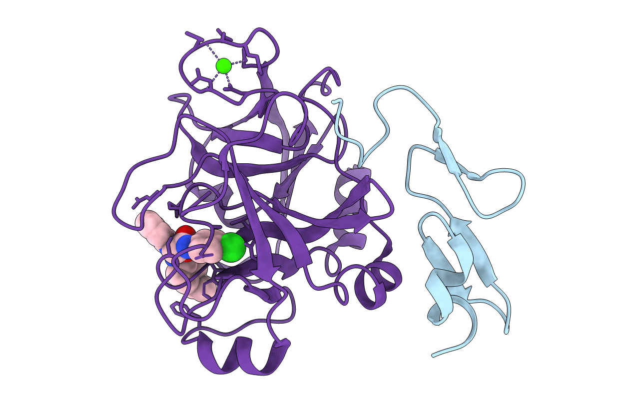
2BQW image
Deposition Date 2005-04-28
Release Date 2006-04-26
Last Version Date 2024-10-16
Entry Detail
PDB ID:
2BQW
Keywords:
Title:
CRYSTAL STRUCTURE OF FACTOR XA IN COMPLEX WITH COMPOUND 45
Biological Source:
Source Organism:
HOMO SAPIENS (Taxon ID: 9606)
Method Details:
Experimental Method:
Resolution:
2.95 Å
R-Value Free:
0.26
R-Value Work:
0.18
Space Group:
P 21 21 21
Macromolecular Entities
Polymer Type:polypeptide(L)
Molecule:COAGULATION FACTOR X
Gene (Uniprot):F10
Chain IDs:A
Chain Length:52
Number of Molecules:1
Biological Source:HOMO SAPIENS
Polymer Type:polypeptide(L)
Molecule:FACTOR XA
Gene (Uniprot):F10
Chain IDs:B
Chain Length:249
Number of Molecules:1
Biological Source:HOMO SAPIENS
Primary Citation
Probing the Subpockets of Factor Xa Reveals Two Binding Modes for Inhibitors Based on a 2-Carboxyindole Scaffold: A Study Combining Structure-Activity Relationship and X-Ray Crystallography.
J.Med.Chem. 48 4511 ? (2005)
PMID: 15999990 DOI: 10.1021/JM0490540

Abstact

Structure-activity relationships within a series of highly potent 2-carboxyindole-based factor Xa inhibitors incorporating a neutral P1 ligand are described with particular emphasis on the structural requirements for addressing subpockets of the factor Xa enzyme. Interactions with the subpockets were probed by systematic substitution of the 2-carboxyindole scaffold, in combination with privileged P1 and P4 substituents. Combining the most favorable substituents at the indole nucleus led to the discovery of a remarkably potent factor Xa inhibitor displaying a K(i) value of 0.07 nM. X-ray crystallography of inhibitors bound to factor Xa revealed substituent-dependent switching of the inhibitor binding mode and provided a rationale for the SAR obtained. These results underscore the key role played by the P1 ligand not only in determining the binding affinity of the inhibitor by direct interaction but also in modifying the binding mode of the whole scaffold, resulting in a nonlinear SAR.

Legend

Protein

Chemical

Disease