Deposition Date
2005-04-20
Release Date
2006-10-11
Last Version Date
2023-12-13
Entry Detail
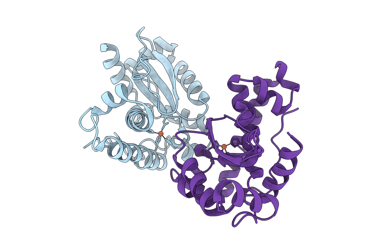
PDB ID:
2BPI
Keywords:
Title:
Structure of Iron dependent superoxide dismutase from P. falciparum.
Biological Source:
Source Organism(s):
PLASMODIUM FALCIPARUM (Taxon ID: 36329)
Expression System(s):
Method Details:
Experimental Method:
Resolution:
2.52 Å
R-Value Free:
0.26
R-Value Work:
0.18
R-Value Observed:
0.18
Space Group:
P 21 21 21