2BP7 image
Deposition Date 2005-04-18
Release Date 2005-08-10
Last Version Date 2023-12-13
Entry Detail
PDB ID:
2BP7
Keywords:
Title:
New crystal form of the Pseudomonas putida branched-chain dehydrogenase (E1)
Biological Source:
Source Organism:
Host Organism:
Method Details:
Experimental Method:
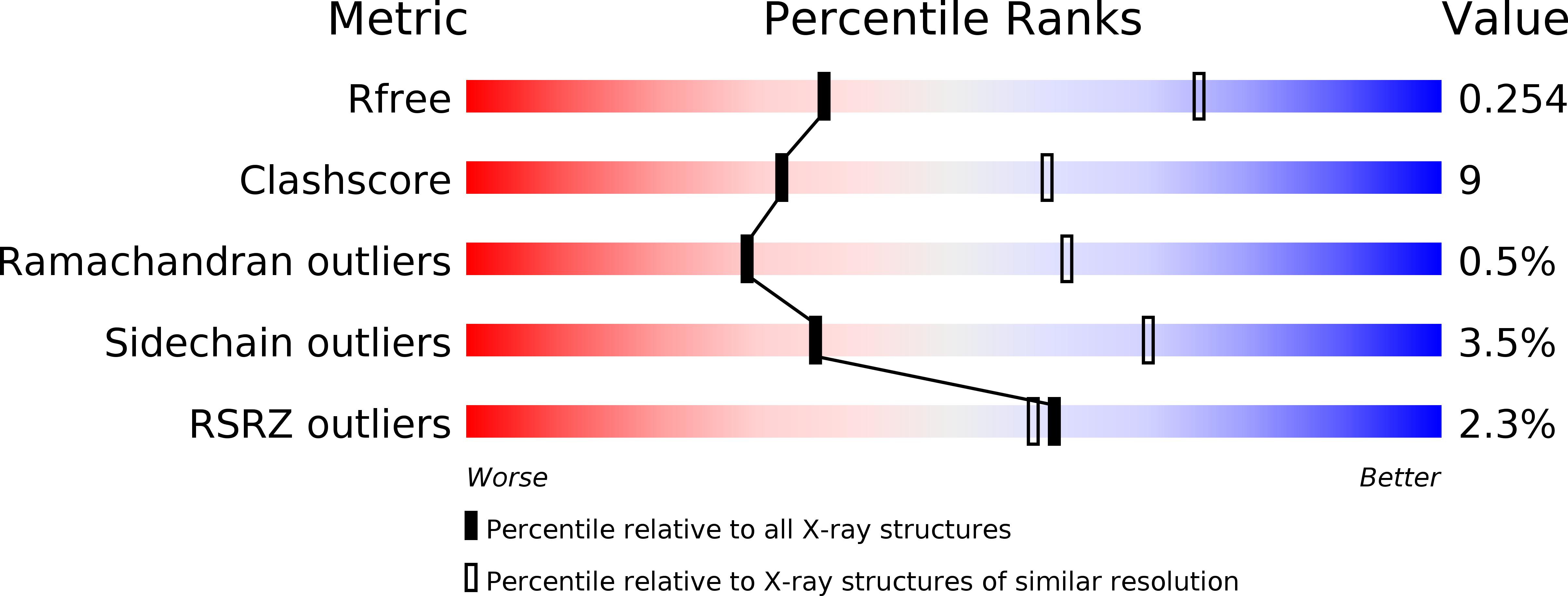
Resolution:
2.90 Å
R-Value Free:
0.25
R-Value Work:
0.22
R-Value Observed:
0.22
Space Group:
P 61 2 2
Macromolecular Entities
Polymer Type:polypeptide(L)
Molecule:2-OXOISOVALERATE DEHYDROGENASE ALPHA SUBUNIT
Gene (Uniprot):bkdA1
Chain IDs:A, C, E, G
Chain Length:410
Number of Molecules:4
Biological Source:PSEUDOMONAS PUTIDA
Polymer Type:polypeptide(L)
Molecule:2-OXOISOVALERATE DEHYDROGENASE BETA SUBUNIT
Gene (Uniprot):bkdA2
Chain IDs:B, D, F, H
Chain Length:339
Number of Molecules:4
Biological Source:PSEUDOMONAS PUTIDA
Primary Citation
The Molecular Origins of Specificity in the Assembly of a Multienzyme Complex.
Structure 13 1119 ? (2005)
PMID: 16084384 DOI: 10.1016/J.STR.2005.04.021

Abstact

The pyruvate dehydrogenase (PDH) multienzyme complex is central to oxidative metabolism. We present the first crystal structure of a complex between pyruvate decarboxylase (E1) and the peripheral subunit binding domain (PSBD) of the dihydrolipoyl acetyltransferase (E2). The interface is dominated by a "charge zipper" of networked salt bridges. Remarkably, the PSBD uses essentially the same zipper to alternately recognize the dihydrolipoyl dehydrogenase (E3) component of the PDH assembly. The PSBD achieves this dual recognition largely through the addition of a network of interfacial water molecules unique to the E1-PSBD complex. These structural comparisons illuminate our observations that the formation of this water-rich E1-E2 interface is largely enthalpy driven, whereas that of the E3-PSBD complex (from which water is excluded) is entropy driven. Interfacial water molecules thus diversify surface complementarity and contribute to avidity, enthalpically. Additionally, the E1-PSBD structure provides insight into the organization and active site coupling within the approximately 9 MDa PDH complex.

Legend

Protein

Chemical

Disease

Primary Citation of related structures