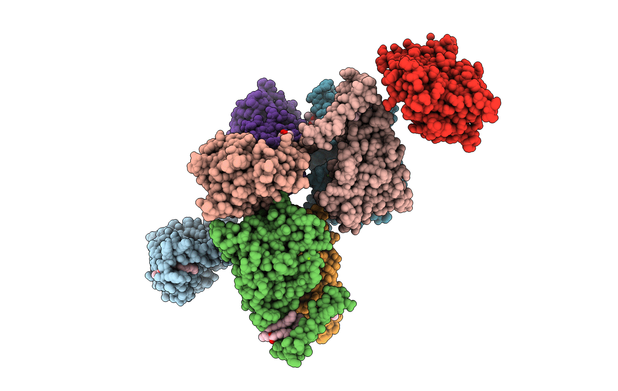
2BOY image
Entry Detail
PDB ID:
2BOY
Keywords:
Title:
Crystal structure of 3-ChloroCatechol 1,2-Dioxygenase from Rhodococcus Opacus 1CP
Biological Source:
Source Organism:
PDB Version:
Deposition Date:
2005-04-15
Release Date:
2006-08-24
Method Details:
Experimental Method:
Resolution:
1.90 Å
R-Value Free:
0.21
R-Value Work:
0.17
R-Value Observed:
0.17
Space Group:
P 1
Macromolecular Entities
Polymer Type:polypeptide(L)
Description:3-CHLOROCATECHOL 1,2-DIOXYGENASE
Chain IDs:A, B, C, D, E, F, G, H
Chain Length:254
Number of Molecules:8
Biological Source:RHODOCOCCUS OPACUS
Primary Citation
Crystal structure of 3-chlorocatechol 1,2-dioxygenase key enzyme of a new modified ortho-pathway from the Gram-positive Rhodococcus opacus 1CP grown on 2-chlorophenol.
J. Mol. Biol. 360 788 799 (2006)
PMID: 16793061 DOI: 10.1016/j.jmb.2006.05.046

Abstact

The crystal structure of the 3-chlorocatechol 1,2-dioxygenase from the Gram-positive bacterium Rhodococcus opacus (erythropolis) 1CP, a Fe(III) ion-containing enzyme specialized in the aerobic biodegradation of 3-chloro- and methyl-substituted catechols, has been solved by molecular replacement techniques using the coordinates of 4-chlorocatechol 1,2-dioxygenase from the same organism (PDB code 1S9A) as a starting model and refined at 1.9 A resolution (R(free) 21.9%; R-factor 17.4%). The analysis of the structure and of the kinetic parameters for a series of different substrates, and the comparison with the corresponding data for the 4-chlorocatechol 1,2-dioxygenase isolated from the same bacterial strain, provides evidence of which active site residues are responsible for the observed differences in substrate specificity. Among the amino acid residues expected to interact with substrates, only three are altered Val53(Ala53), Tyr78(Phe78) and Ala221(Cys224) (3-chlorocatechol 1,2-dioxygenase(4-chlorocatechol 1,2-dioxygenase)), clearly identifying the substitutions influencing substrate selectivity in these enzymes. The crystallographic asymmetric unit contains eight subunits (corresponding to four dimers) that show heterogeneity in the conformation of a co-crystallized molecule bound to the catalytic non-heme iron(III) ion resembling a benzohydroxamate moiety, probably a result of the breakdown of recently discovered siderophores synthesized by Gram-positive bacteria. Several different modes of binding benzohydroxamate into the active site induce distinct conformations of the interacting protein ligands Tyr167 and Arg188, illustrating the plasticity of the active site origin of the more promiscuous substrate preferences of the present enzyme.

Legend

Protein

Chemical

Disease

Primary Citation of related structures