2BOE image
Entry Detail
PDB ID:
2BOE
Keywords:
Title:
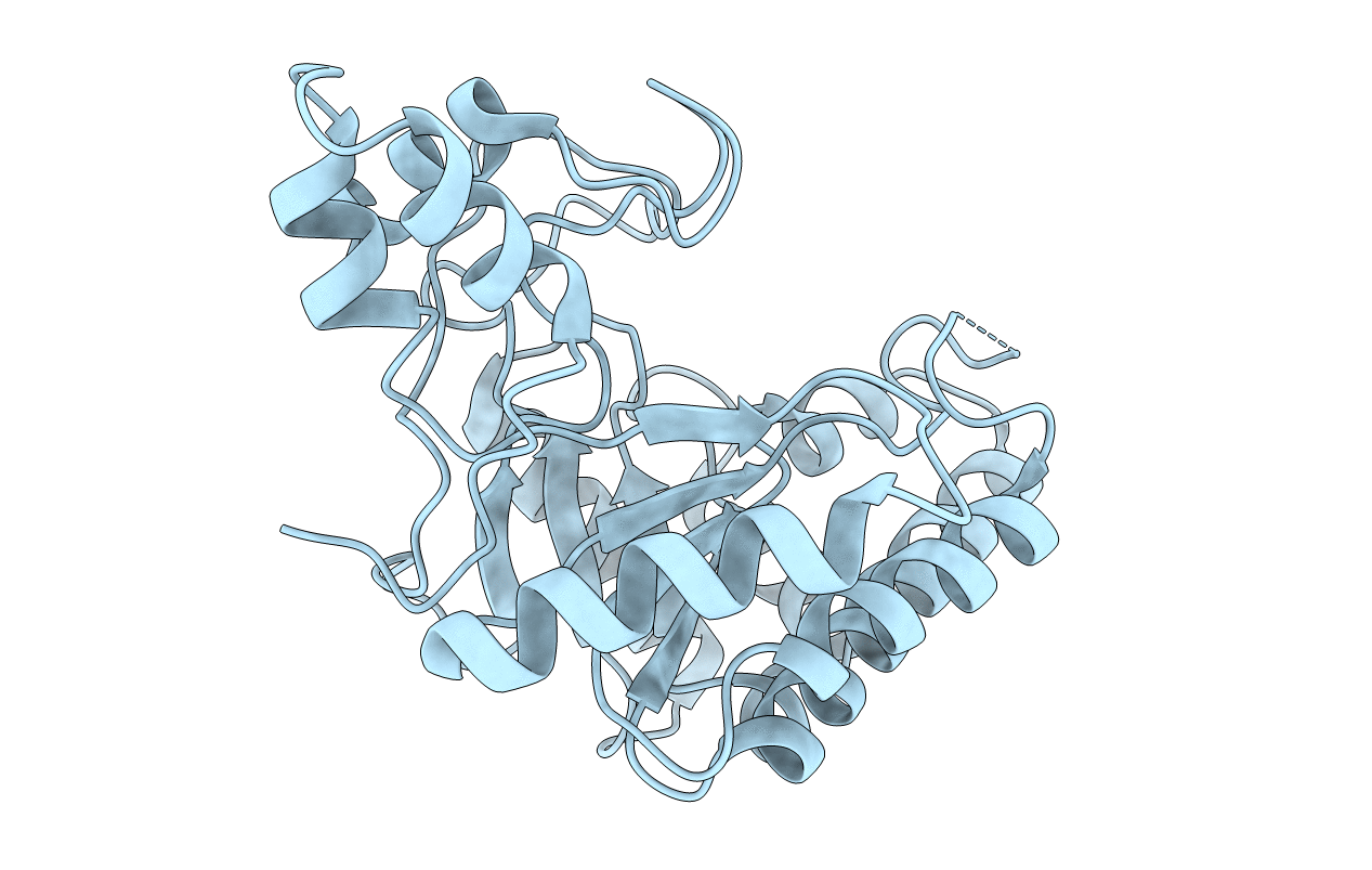
Catalytic domain of endo-1,4-glucanase Cel6A mutant Y73S from Thermobifida fusca
Biological Source:
Source Organism:
Host Organism:
PDB Version:
Deposition Date:
2005-04-10
Release Date:
2005-10-05
Method Details:
Experimental Method:
Resolution:
1.15 Å
R-Value Free:
0.19
R-Value Work:
0.16
R-Value Observed:
0.16
Space Group:
P 21 21 21
Macromolecular Entities
Polymer Type:polypeptide(L)
Description:ENDOGLUCANASE E-2
Mutations:YES
Chain IDs:A (auth: X)
Chain Length:286
Number of Molecules:1
Biological Source:THERMOMONOSPORA FUSCA
Primary Citation
Crystal Structure of Thermobifida Fusca Endoglucanase Cel6A in Complex with Substrate and Inhibitor: The Role of Tyrosine Y73 in Substrate Ring Distortion.
Biochemistry 44 12915 ? (2005)
PMID: 16185060 DOI: 10.1021/BI0506730

Abstact

Endoglucanase Cel6A from Thermobifida fusca hydrolyzes the beta-1,4 linkages in cellulose at accessible points along the polymer. The structure of the catalytic domain of Cel6A from T. fusca in complex with a nonhydrolysable substrate analogue that acts as an inhibitor, methylcellobiosyl-4-thio-beta-cellobioside (Glc(2)-S-Glc(2)), has been determined to 1.5 A resolution. The glycosyl unit in subsite -1 was sterically hindered by Tyr73 and forced into a distorted (2)S(o) conformation. In the enzyme where Tyr73 was mutated to a serine residue, the hindrance was removed and the glycosyl unit in subsite -1 had a relaxed (4)C(1) chair conformation. The relaxed conformation was seen in two complex structures of the mutated enzyme, with cellotetrose (Glc(4)) at 1.64 A and Glc(2)-S-Glc(2) at 1.04 A resolution.

Legend

Protein

Chemical

Disease

Primary Citation of related structures