2BNZ image
Deposition Date 2005-04-06
Release Date 2006-03-15
Last Version Date 2023-12-13
Entry Detail
PDB ID:
2BNZ
Title:
Structural basis for cooperative binding of Ribbon-Helix-Helix Omega repressor to inverted DNA heptad repeats
Biological Source:
Source Organism:
Host Organism:
Method Details:
Experimental Method:
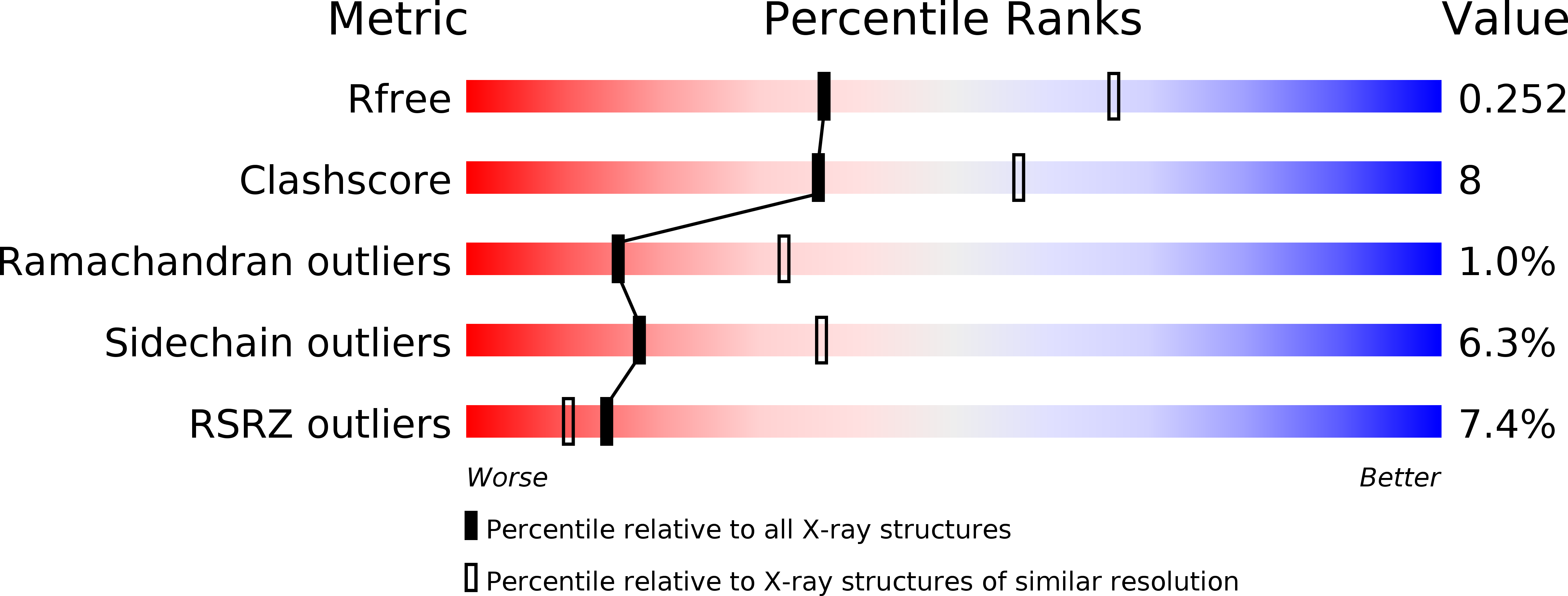
Resolution:
2.60 Å
R-Value Free:
0.25
R-Value Work:
0.22
R-Value Observed:
0.22
Space Group:
P 1 21 1
Macromolecular Entities
Structures with similar UniProt ID
Protein Blast
Polymer Type:polypeptide(L)
Molecule:ORF OMEGA
Gene (Uniprot):omega
Chain IDs:A, B, C, D
Chain Length:53
Number of Molecules:4
Biological Source:STREPTOCOCCUS PYOGENES
Polymer Type:polydeoxyribonucleotide
Molecule:5'-D(*GP*AP*AP*TP*CP*AP*CP*AP*AP*GP *TP*GP*AP*TP*TP*AP*GP*C)-3'
Chain IDs:E, G
Chain Length:18
Number of Molecules:2
Biological Source:SYNTHETIC CONSTRUCT
Polymer Type:polydeoxyribonucleotide
Molecule:5'-D(*CP*TP*AP*AP*TP*CP*AP*CP*TP*TP *GP*TP*GP*AP*TP*TP*CP*G)-3'
Chain IDs:F, H
Chain Length:18
Number of Molecules:2
Biological Source:SYNTHETIC CONSTRUCT
Primary Citation
Structures of Omega Repressors Bound to Direct and Inverted DNA Repeats Explain Modulation of Transcription.
Nucleic Acids Res. 34 1450 ? (2006)
PMID: 16528102 DOI: 10.1093/NAR/GKL015

Abstact

Repressor omega regulates transcription of genes required for copy number control, accurate segregation and stable maintenance of inc18 plasmids hosted by Gram-positive bacteria. omega belongs to homodimeric ribbon-helix-helix (RHH2) repressors typified by a central, antiparallel beta-sheet for DNA major groove binding. Homodimeric omega2 binds cooperatively to promotors with 7 to 10 consecutive non-palindromic DNA heptad repeats (5'-(A)/(T)ATCAC(A)/(T)-3', symbolized by -->) in palindromic inverted, converging (--><--) or diverging (<---->) orientation and also, unique to omega2 and contrasting other RHH2 repressors, to non-palindromic direct (-->-->) repeats. Here we investigate with crystal structures how omega2 binds specifically to heptads in minimal operators with (-->-->) and (--><--) repeats. Since the pseudo-2-fold axis relating the monomers in omega(2) passes the central C-G base pair of each heptad with approximately 0.3 A downstream offset, the separation between the pseudo-2-fold axes is exactly 7 bp in (-->-->), approximately 0.6 A shorter in (--><--) but would be approximately 0.6 A longer in (<---->). These variations grade interactions between adjacent omega2 and explain modulations in cooperative binding affinity of omega2 to operators with different heptad orientations.

Legend

Protein

Chemical

Disease

Primary Citation of related structures