Deposition Date
2005-02-23
Release Date
2005-10-13
Last Version Date
2024-05-08
Entry Detail
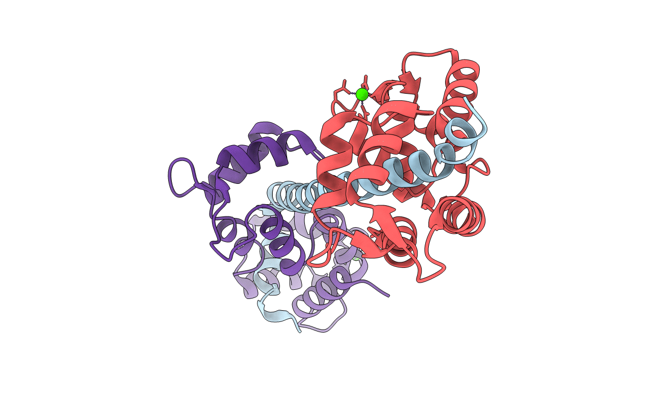
Biological Source:
Source Organism(s):
PHYSARUM POLYCEPHALUM (Taxon ID: 5791)
Expression System(s):
Method Details:
Experimental Method:
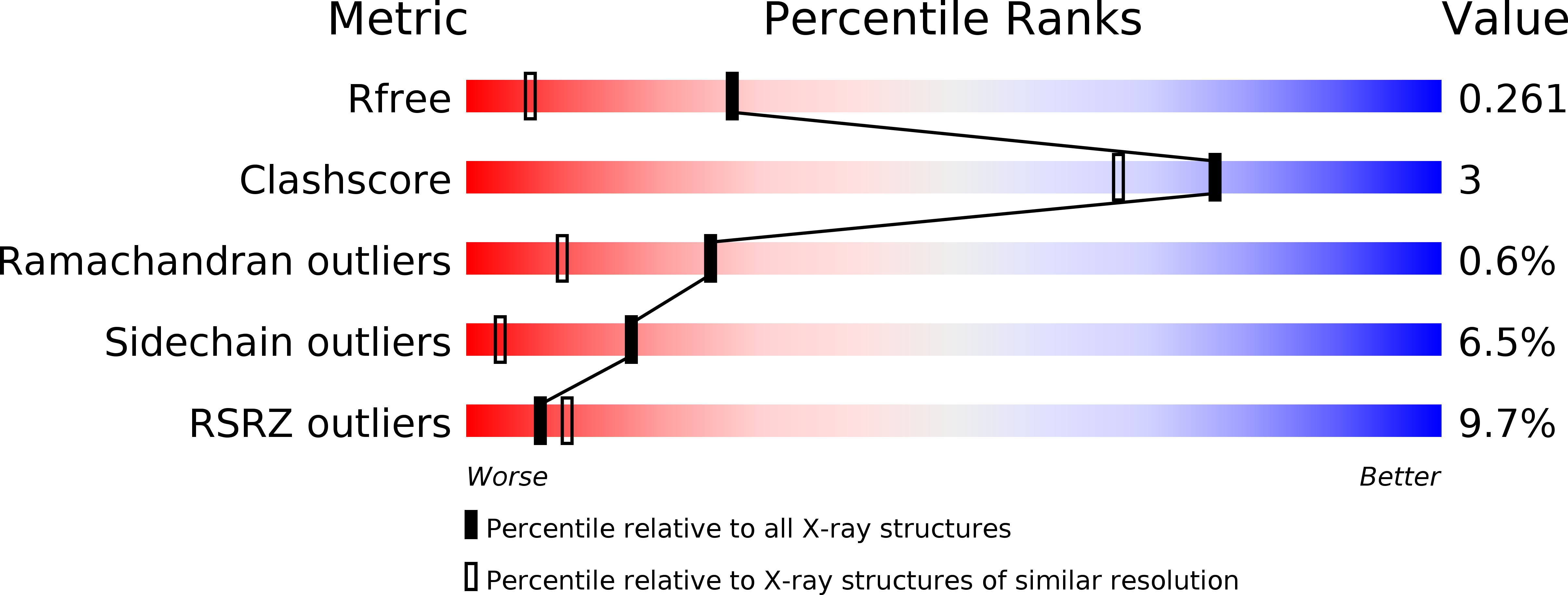
Resolution:
1.75 Å
R-Value Free:
0.25
R-Value Work:
0.21
R-Value Observed:
0.22
Space Group:
P 21 21 21