2BKM image
Entry Detail
PDB ID:
2BKM
Keywords:
Title:
Crystal structure of the truncated hemoglobin from Geobacillus stearothermophilus
Biological Source:
PDB Version:
Deposition Date:
2006-02-08
Release Date:
2006-11-29
Method Details:
Experimental Method:
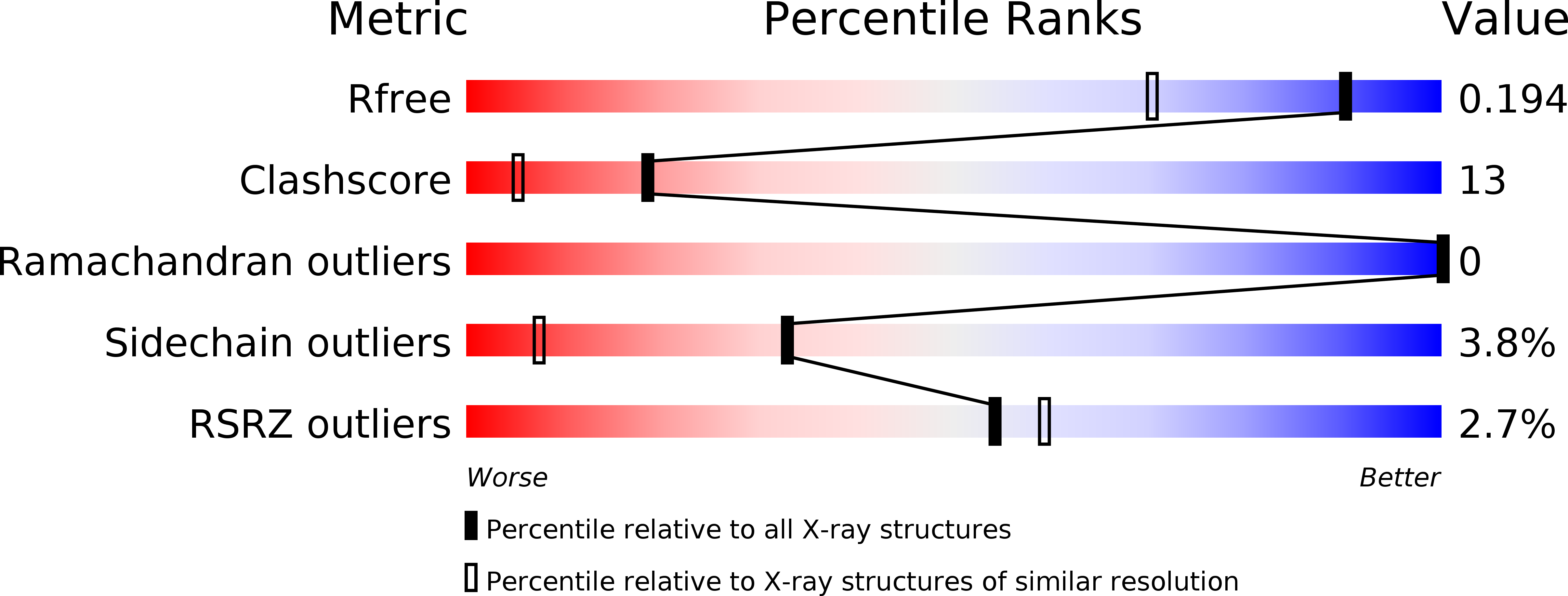
Resolution:
1.50 Å
R-Value Free:
0.19
R-Value Work:
0.16
R-Value Observed:
0.16
Space Group:
P 1 21 1
Macromolecular Entities
Polymer Type:polypeptide(L)
Description:TRUNCATED HEMOGLOBIN FROM GEOBACILLUS STEAROTHERMOPHILUS
Chain IDs:A, B
Chain Length:128
Number of Molecules:2
Biological Source:GEOBACILLUS STEAROTHERMOPHILUS
Primary Citation
Crystal Structure and Ligand Binding Properties of the Truncated Hemoglobin from Geobacillus Stearothermophilus
Arch.Biochem.Biophys. 457 85 ? (2007)
PMID: 17126283 DOI: 10.1016/J.ABB.2006.09.033

Abstact

A novel truncated hemoglobin has been identified in the thermophilic bacterium Geobacillus stearothermophilus (Gs-trHb). The protein has been expressed in Escherichia coli, the 3D crystal structure (at 1.5 Angstroms resolution) and the ligand binding properties have been determined. The distal heme pocket displays an array of hydrogen bonding donors to the iron-bound ligands, including Tyr-B10 on one side of the heme pocket and Trp-G8 indole nitrogen on the opposite side. At variance with the highly similar Bacillus subtilis hemoglobin, Gs-trHb is dimeric both in the crystal and in solution and displays several unique structural properties. In the crystal cell, the iron-bound ligand is not homogeneously distributed within each distal site such that oxygen and an acetate anion can be resolved with relative occupancies of 50% each. Accordingly, equilibrium titrations of the oxygenated derivative in solution with acetate anion yield a partially saturated ferric acetate adduct. Moreover, the asymmetric unit contains two subunits and sedimentation velocity ultracentrifugation data confirm that the protein is dimeric.

Legend

Protein

Chemical

Disease

Primary Citation of related structures