2BKG image
Entry Detail
PDB ID:
2BKG
Keywords:
Title:
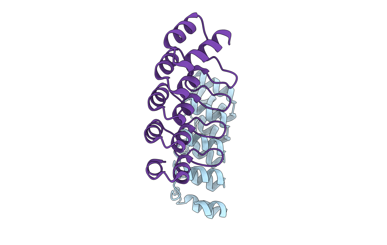
Crystal structure of E3_19 a designed ankyrin repeat protein
Biological Source:
Source Organism:
Host Organism:
PDB Version:
Deposition Date:
2005-02-16
Release Date:
2006-06-21
Method Details:
Experimental Method:
Resolution:
1.90 Å
R-Value Free:
0.22
R-Value Work:
0.17
R-Value Observed:
0.17
Space Group:
P 21 21 21
Macromolecular Entities
Polymer Type:polypeptide(L)
Description:SYNTHETIC CONSTRUCT ANKYRIN REPEAT PROTEIN E3_19
Chain IDs:A, B
Chain Length:166
Number of Molecules:2
Biological Source:SYNTHETIC CONSTRUCT
Primary Citation
Crystal Structure of a Consensus-Designed Ankyrin Repeat Protein: Implications for Stability
Proteins: Struct., Funct., Bioinf. 65 280 ? (2006)
PMID: 16493627 DOI: 10.1002/PROT.20930

Abstact

Consensus-designed ankyrin repeat (AR) proteins are thermodynamically very stable. The structural analysis of the designed AR protein E3_5 revealed that this stability is due to a regular fold with highly conserved structural motifs and H-bonding networks. However, the designed AR protein E3_19 exhibits a significantly lower stability than E3_5 (9.6 vs. 14.8 kcal/mol), despite 88% sequence identity. To investigate the structural correlations of this stability difference between E3_5 and E3_19, we determined the crystal structure of E3_19 at 1.9 A resolution. E3_19 as well has a regular AR domain fold with the characteristic H-bonding patterns. All structural features of the E3_5 and E3_19 molecules appear to be virtually identical (RMSD(Calpha) approximately 0.7 A). However, clear differences are observed in the surface charge distribution of the two AR proteins. E3_19 features clusters of charged residues and more exposed hydrophobic residues than E3_5. The atomic coordinates of E3_19 have been deposited in the Protein Data Bank. PDB ID: 2BKG.

Legend

Protein

Chemical

Disease

Primary Citation of related structures