Deposition Date
2005-02-15
Release Date
2005-03-16
Last Version Date
2024-10-09
Entry Detail
PDB ID:
2BKE
Keywords:
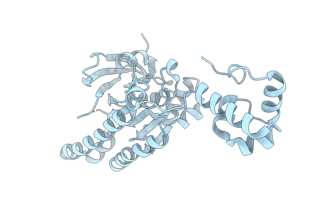
Title:
Conformational Flexibility Revealed by the Crystal Structure of a Crenarchaeal RadA
Biological Source:
Source Organism(s):
SULFOLOBUS SOLFATARICUS (Taxon ID: 273057)
Expression System(s):
Method Details:
Experimental Method:
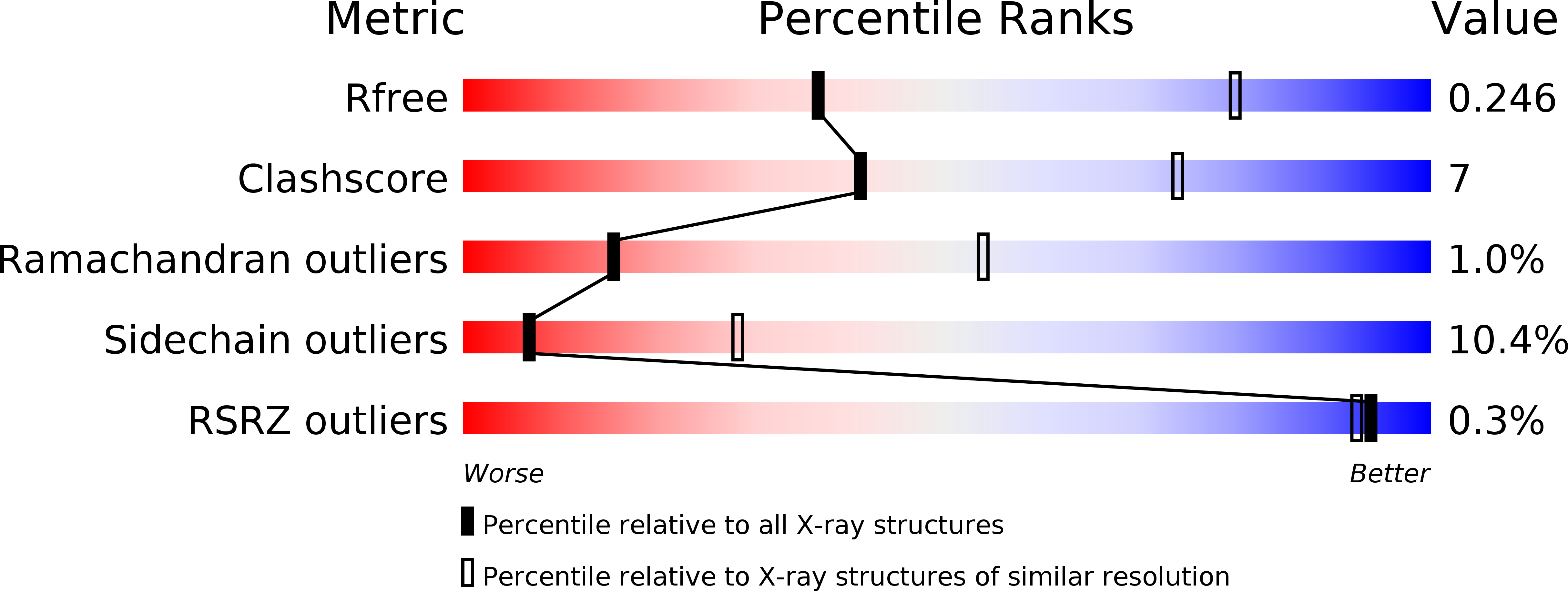
Resolution:
3.20 Å
R-Value Free:
0.24
R-Value Work:
0.17
R-Value Observed:
0.17
Space Group:
P 31 2 1