Deposition Date
2005-01-26
Release Date
2005-04-14
Last Version Date
2024-05-08
Entry Detail
PDB ID:
2BIX
Keywords:
Title:
Crystal structure of apocarotenoid cleavage oxygenase from Synechocystis, Fe-free apoenzyme
Biological Source:
Source Organism(s):
SYNECHOCYSTIS SP. (Taxon ID: 1148)
Expression System(s):
Method Details:
Experimental Method:
Resolution:
2.68 Å
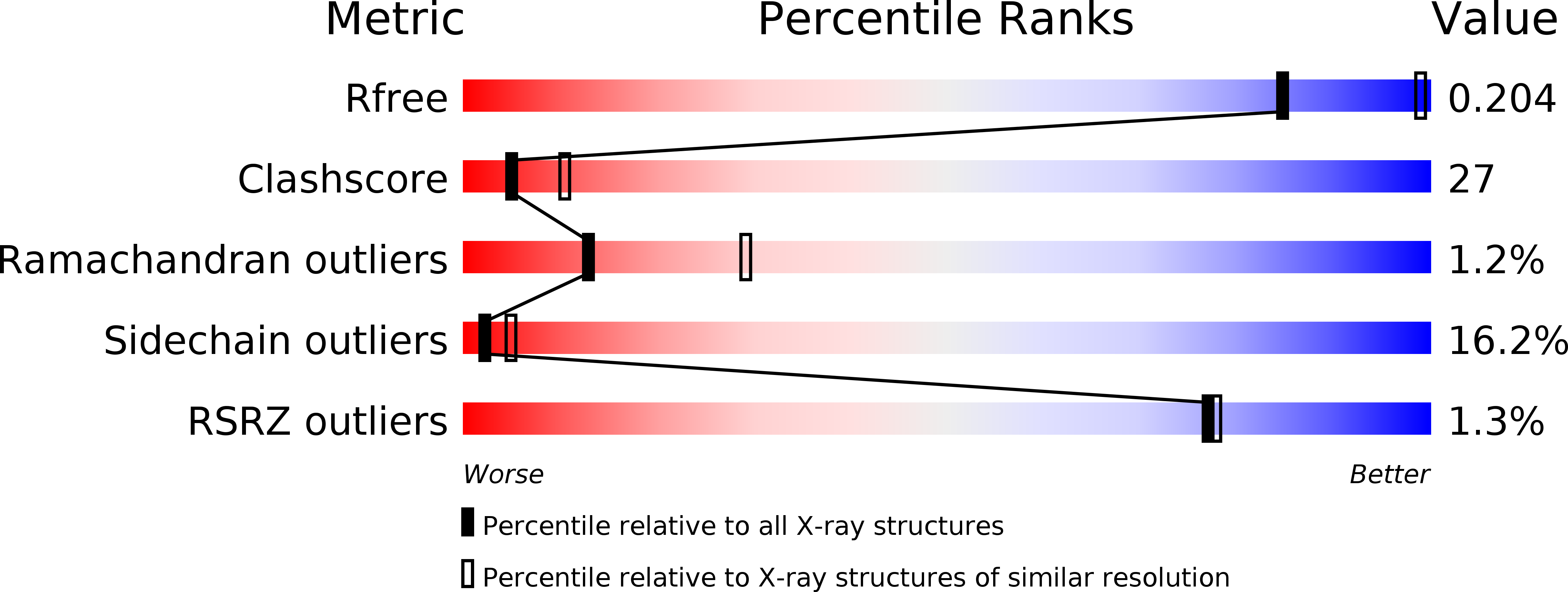
R-Value Free:
0.24
R-Value Work:
0.21
R-Value Observed:
0.21
Space Group:
P 41 21 2