2BIW image
Deposition Date 2005-01-26
Release Date 2005-04-14
Last Version Date 2024-05-01
Entry Detail
PDB ID:
2BIW
Keywords:
Title:
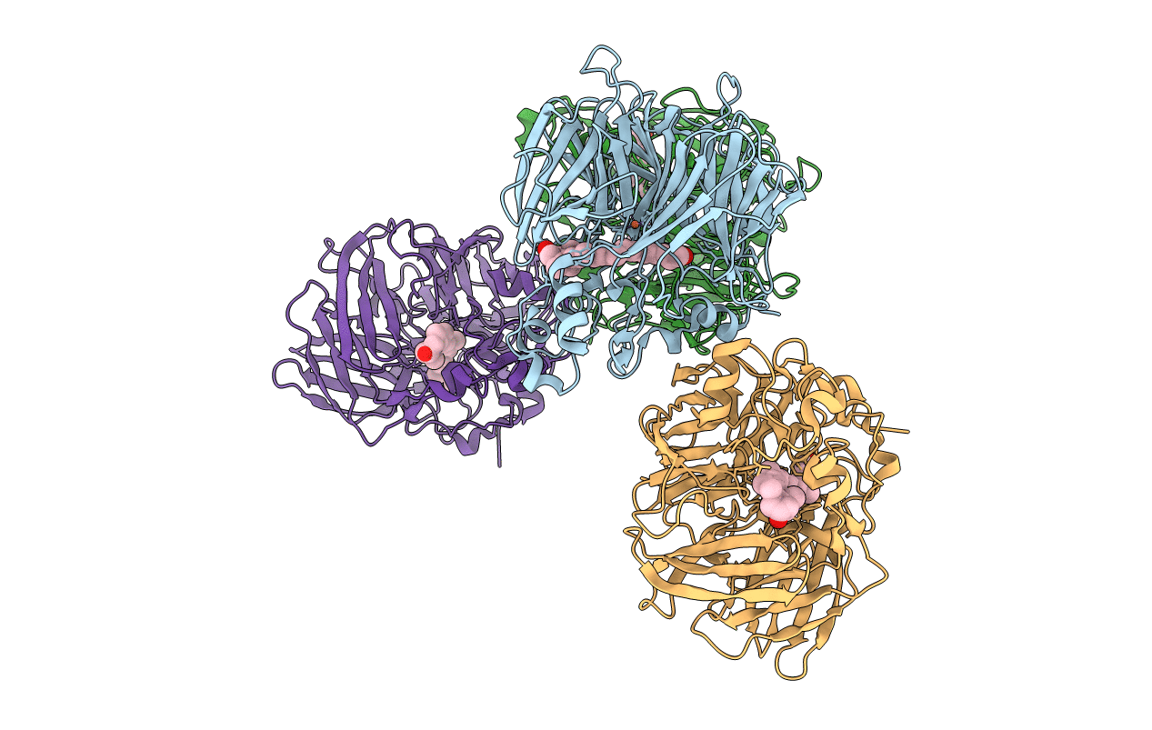
Crystal structure of apocarotenoid cleavage oxygenase from Synechocystis, native enzyme
Biological Source:
Source Organism:
Host Organism:
Method Details:
Experimental Method:
Resolution:
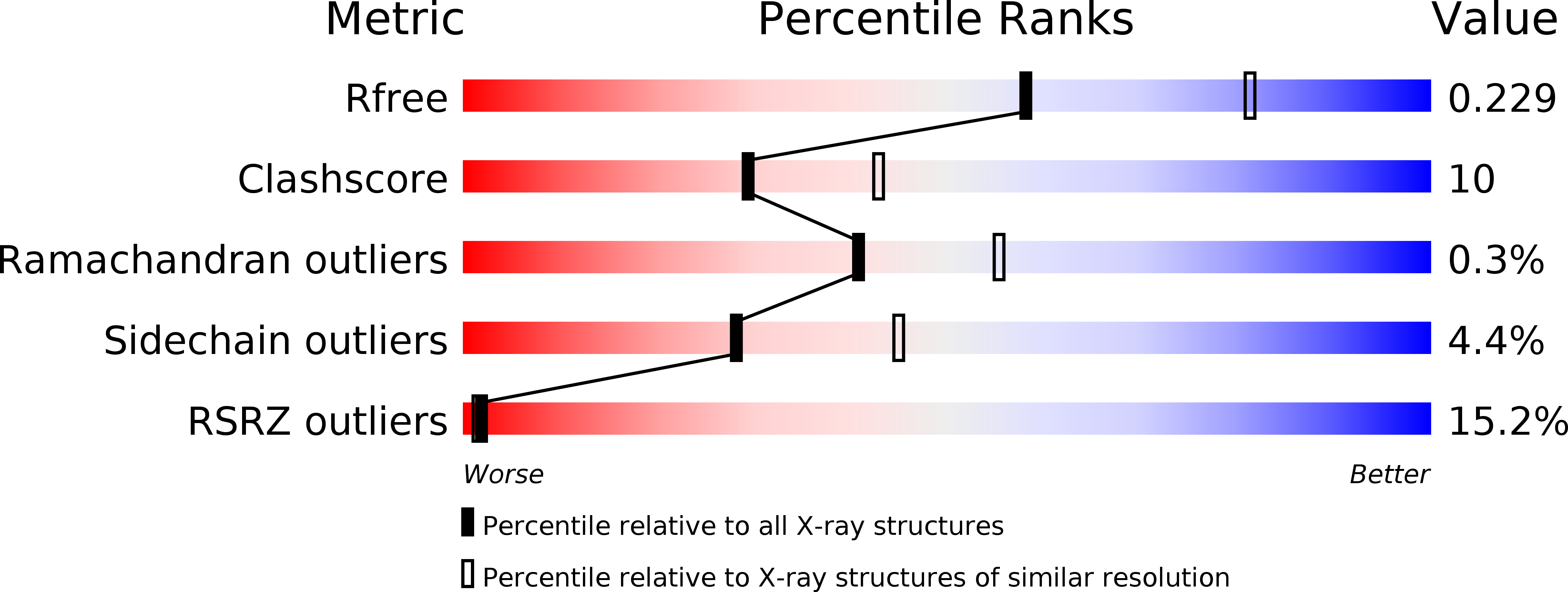
2.39 Å
R-Value Free:
0.22
R-Value Work:
0.18
R-Value Observed:
0.18
Space Group:
P 21 21 21
Macromolecular Entities
Structures with similar UniProt ID
Protein Blast
Polymer Type:polypeptide(L)
Molecule:APOCAROTENOID-CLEAVING OXYGENASE
Gene (Uniprot):sll1541
Chain IDs:A, B, C, D
Chain Length:490
Number of Molecules:4
Biological Source:SYNECHOCYSTIS SP.
Primary Citation
The Structure of a Retinal-Forming Carotenoid Oxygenase
Science 308 267 ? (2005)
PMID: 15821095 DOI: 10.1126/SCIENCE.1108965

Abstact

Enzymes that produce retinal and related apocarotenoids constitute a sequence- and thus structure-related family, a member of which was analyzed by x-ray diffraction. This member is an oxygenase and contains an Fe2+-4-His arrangement at the axis of a seven-bladed beta-propeller chain fold covered by a dome formed by six large loops. The Fe2+ is accessible through a long nonpolar tunnel that holds a carotenoid derivative in one of the crystals. On binding, three consecutive double bonds of this carotenoid changed from a straight all-trans to a cranked cis-trans-cis conformation. The remaining trans bond is located at the dioxygen-ligated Fe2+ and cleaved by oxygen.

Legend

Protein

Chemical

Disease

Primary Citation of related structures