Deposition Date
2005-01-26
Release Date
2005-01-26
Last Version Date
2023-12-13
Entry Detail
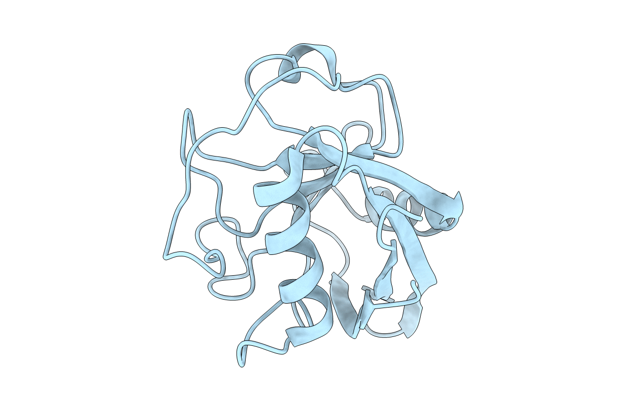
PDB ID:
2BIT
Keywords:
Title:
Crystal structure of human cyclophilin D at 1.7 A resolution
Biological Source:
Source Organism(s):
HOMO SAPIENS (Taxon ID: 9606)
Expression System(s):
Method Details:
Experimental Method:
Resolution:
1.71 Å
R-Value Free:
0.17
R-Value Work:
0.13
R-Value Observed:
0.14
Space Group:
P 41 21 2