Deposition Date
2005-01-25
Release Date
2005-01-26
Last Version Date
2023-12-13
Entry Detail
PDB ID:
2BIO
Keywords:
Title:
human p53 core domain mutant M133L-V203A-N239Y-R249S-N268D
Biological Source:
Source Organism(s):
HOMO SAPIENS (Taxon ID: 9606)
Expression System(s):
Method Details:
Experimental Method:
Resolution:
1.90 Å
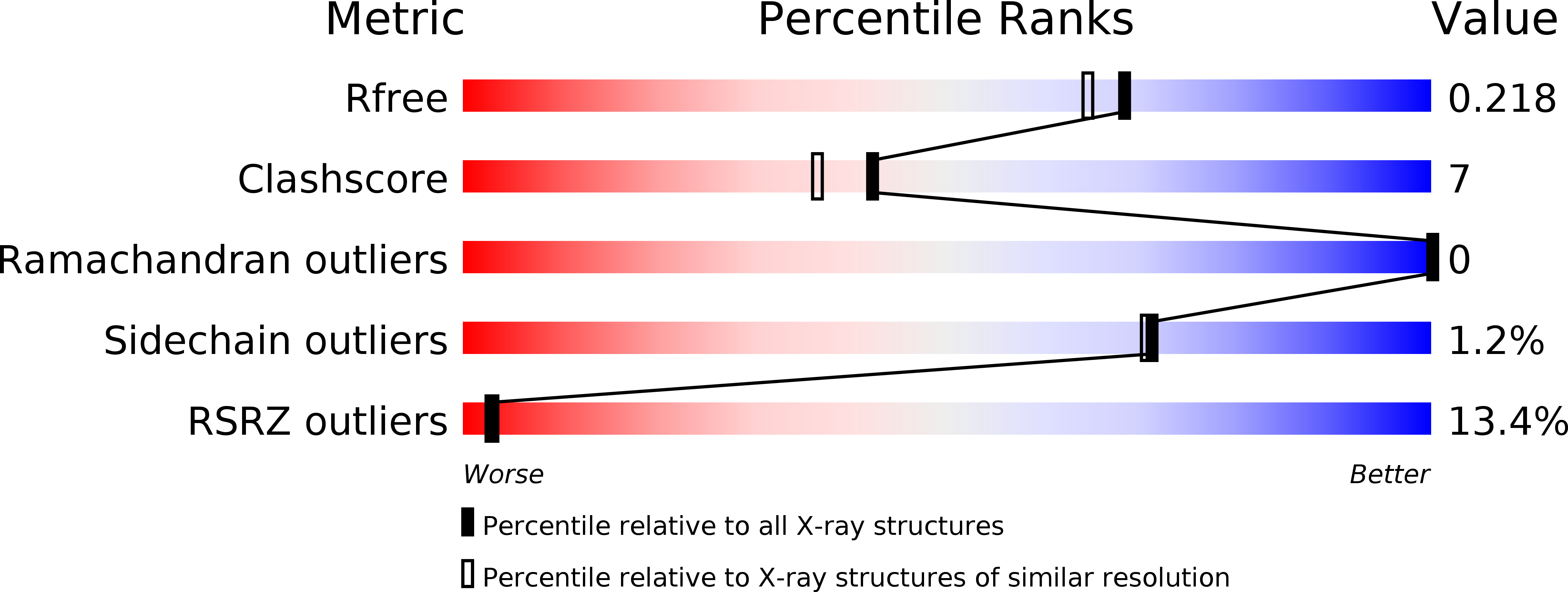
R-Value Free:
0.23
R-Value Work:
0.21
R-Value Observed:
0.21
Space Group:
P 65 2 2