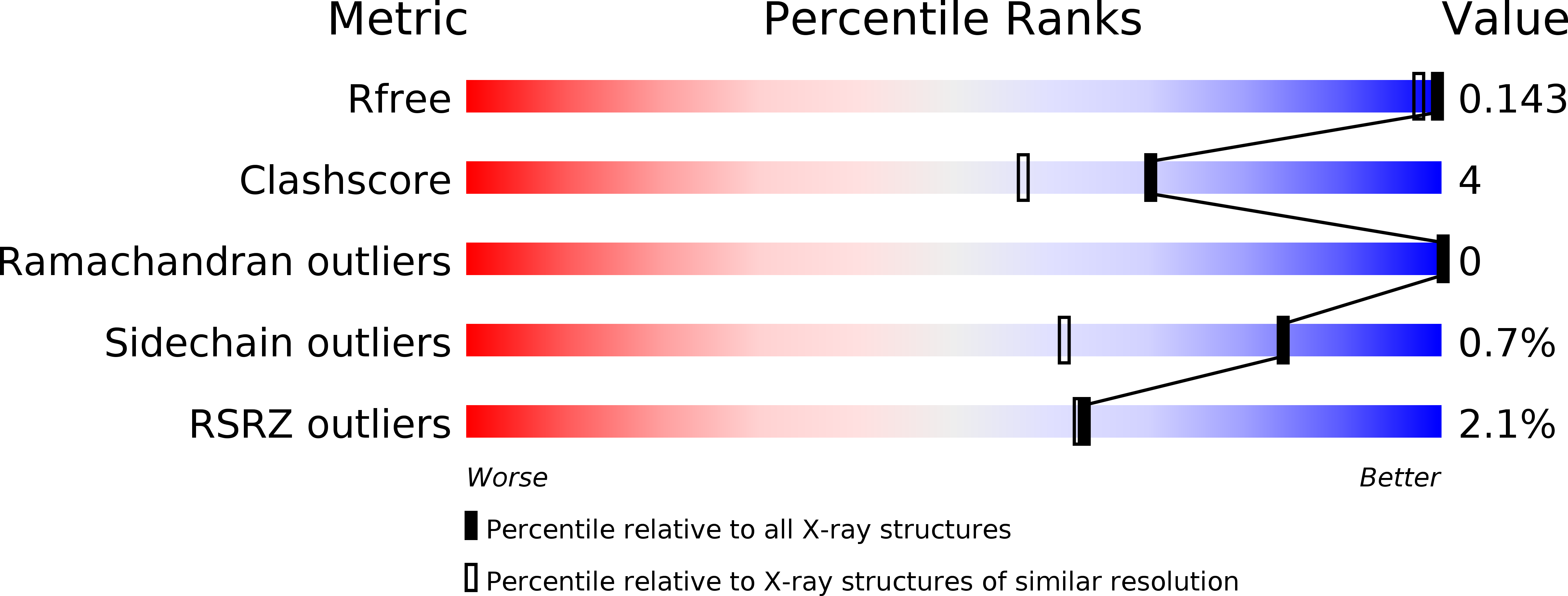
2BIE image
Deposition Date 2005-01-21
Release Date 2005-05-19
Last Version Date 2025-04-09
Entry Detail
PDB ID:
2BIE
Keywords:
Title:
Radiation damage of the Schiff base in phosphoserine aminotransferase (structure H)
Biological Source:
Source Organism:
Host Organism:
Method Details:
Experimental Method:
Resolution:
1.30 Å
R-Value Free:
0.16
R-Value Observed:
0.12
Space Group:
P 21 21 2
Macromolecular Entities
Polymer Type:polypeptide(L)
Molecule:PHOSPHOSERINE AMINOTRANSFERASE
Gene (Uniprot):serC
Chain IDs:A, B
Chain Length:361
Number of Molecules:2
Biological Source:BACILLUS ALCALOPHILUS
Primary Citation
Strain Relief at the Active Site of Phosphoserine Aminotransferase Induced by Radiation Damage.
Protein Sci. 14 1498 ? (2005)
PMID: 15883191 DOI: 10.1110/PS.051397905

Abstact

The X-ray susceptibility of the lysine-pyridoxal-5'-phosphate Schiff base in Bacillus alcalophilus phosphoserine aminotransferase has been investigated using crystallographic data collected at 100 K to 1.3 A resolution, complemented by on-line spectroscopic studies. X-rays induce deprotonation of the internal aldimine, changes in the Schiff base conformation, displacement of the cofactor molecule, and disruption of the Schiff base linkage between pyridoxal-5'-phosphate and the Lys residue. Analysis of the "undamaged" structure reveals a significant chemical strain on the internal aldimine bond that leads to a pronounced geometrical distortion of the cofactor. However, upon crystal exposure to the X-rays, the strain and distortion are relaxed and eventually diminished when the total absorbed dose has exceeded 4.7 x 10(6) Ggamma. Our data provide new insights into the enzymatic activation of pyridoxal-5'-phosphate and suggest that special care should be taken while using macromolecular crystallography to study details in strained active sites.

Legend

Protein

Chemical

Disease

Primary Citation of related structures