Deposition Date
2005-01-20
Release Date
2005-05-19
Last Version Date
2025-04-09
Entry Detail
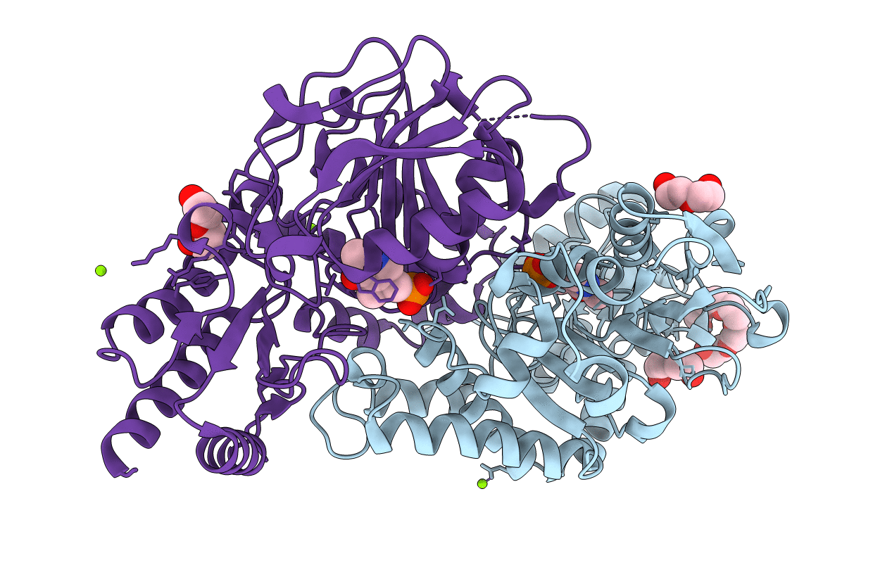
PDB ID:
2BI3
Keywords:
Title:
Radiation damage of the Schiff base in phosphoserine aminotransferase (structure D)
Biological Source:
Source Organism(s):
BACILLUS ALCALOPHILUS (Taxon ID: 1445)
Expression System(s):
Method Details:
Experimental Method:
Resolution:
1.69 Å
R-Value Free:
0.22
R-Value Observed:
0.17
Space Group:
P 21 21 2