2BHY image
Deposition Date 2005-01-20
Release Date 2005-03-31
Last Version Date 2025-10-01
Entry Detail
PDB ID:
2BHY
Keywords:
Title:
Crystal structure of Deinococcus radiodurans maltooligosyltrehalose trehalohydrolase in complex with trehalose
Biological Source:
Source Organism:
Host Organism:
Method Details:
Experimental Method:
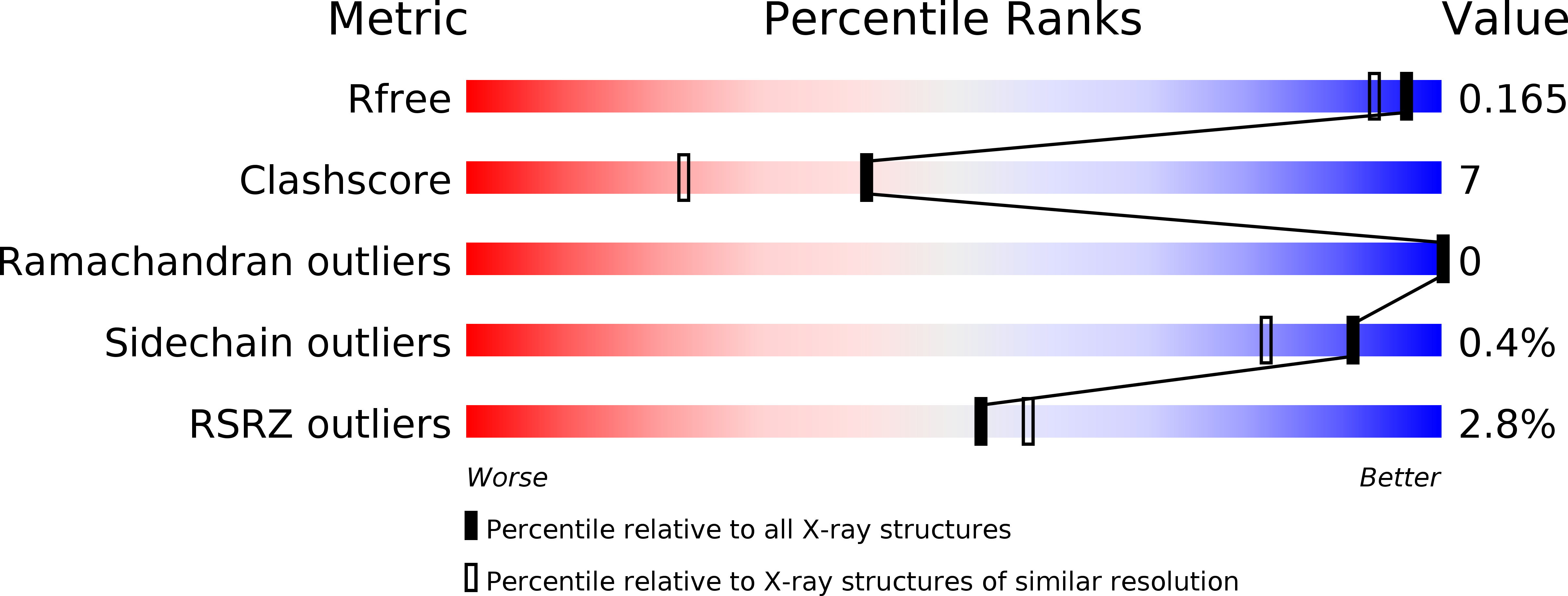
Resolution:
1.50 Å
R-Value Free:
0.15
R-Value Work:
0.12
R-Value Observed:
0.12
Space Group:
P 21 21 21
Macromolecular Entities
Structures with similar UniProt ID
Protein Blast
Polymer Type:polypeptide(L)
Molecule:MALTOOLIGOSYLTREHALOSE TREHALOHYDROLASE
Gene (Uniprot):treZ
Mutagens:YES
Chain IDs:A
Chain Length:602
Number of Molecules:1
Biological Source:DEINOCOCCUS RADIODURANS
Modified Residue
Compound ID Chain ID Parent Comp ID Details 2D Image
MSE A MET SELENOMETHIONINE
Peptide-like Molecules
PRD_900006
Primary Citation
Crystal Structure of Maltooligosyltrehalose Trehalohydrolase from Deinococcus Radiodurans in Complex with Disaccharides
J.Mol.Biol. 347 949 ? (2005)
PMID: 15784255 DOI: 10.1016/J.JMB.2005.02.011

Abstact

Trehalose (alpha-D-glucopyranosyl-1,1-alpha-D-glucopyranose) is a non-reducing diglucoside found in various organisms that serves as a carbohydrate reserve and as an agent that protects against a variety of physical and chemical stresses. Deinococcus radiodurans possesses an alternative biosynthesis pathway for the synthesis of trehalose from maltooligosaccharides. This reaction is mediated by two enzymes: maltooligosyltrehalose synthase (MTSase) and maltooligosyltrehalose trehalohydrolase (MTHase). Here, we present the 1.1A resolution crystal structure of MTHase. It consists of three major domains: two beta-sheet domains and a conserved glycosidase (beta/alpha)8 barrel catalytic domain. Three subdomains consisting of short insertions were identified within the catalytic domain. Subsequently, structures of MTHase in complex with maltose and trehalose were obtained at 1.2 A and 1.5 A resolution, respectively. These structures reveal the importance of the three inserted subdomains in providing the key residues required for substrate recognition. Trehalose is recognised specifically in the +1 and +2 binding subsites by an extensive hydrogen-bonding network and a strong hydrophobic stacking interaction in between two aromatic residues. Moreover, upon binding to maltose, which mimics the substrate sugar chain, a major concerted conformational change traps the sugar chain in the active site. The presence of magnesium in the active site of the MTHase-maltose complex suggests that MTHase activity may be regulated by divalent cations.

Legend

Protein

Chemical

Disease

Primary Citation of related structures