2BHS image
Deposition Date 2005-01-18
Release Date 2005-06-22
Last Version Date 2025-04-09
Entry Detail
PDB ID:
2BHS
Keywords:
Title:
Crystal Structure of Cysteine Synthase B
Biological Source:
Source Organism:
ESCHERICHIA COLI (Taxon ID: 562)
Host Organism:
Method Details:
Experimental Method:
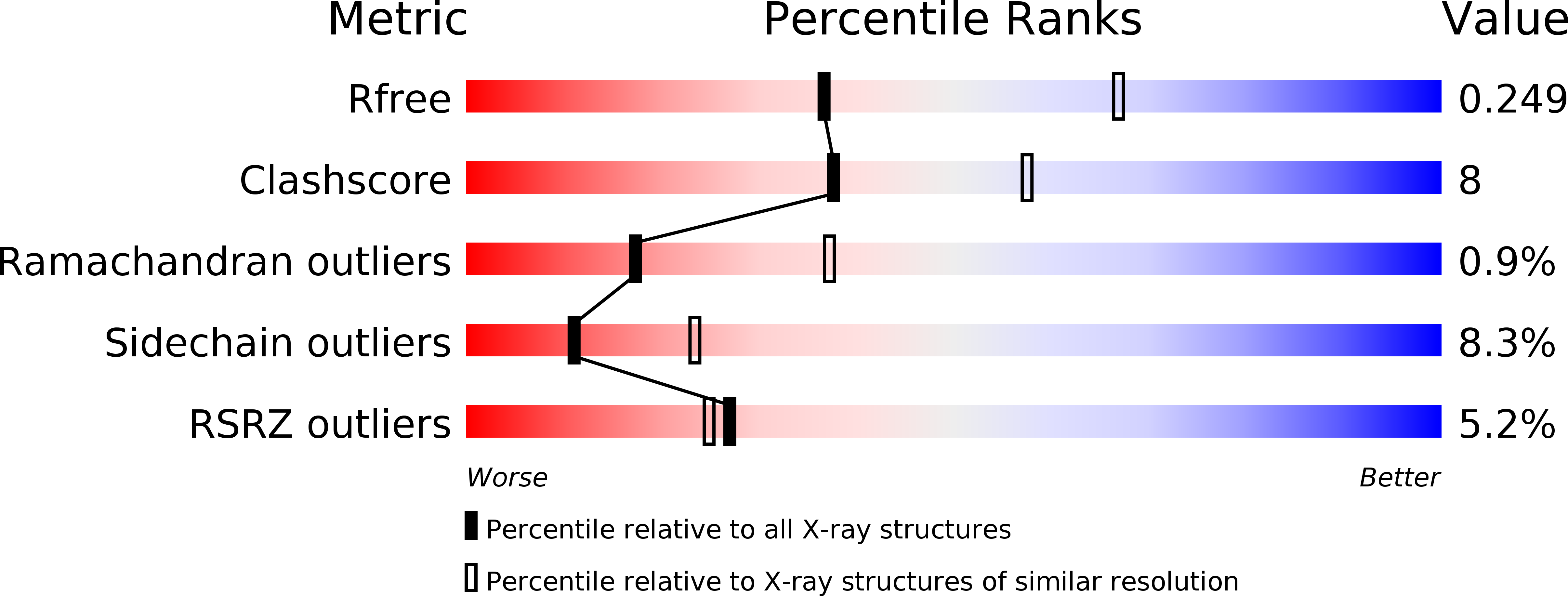
Resolution:
2.67 Å
R-Value Free:
0.25
R-Value Work:
0.22
R-Value Observed:
0.22
Space Group:
P 65 2 2
Macromolecular Entities
Structures with similar UniProt ID
Protein Blast
Polymer Type:polypeptide(L)
Molecule:CYSTEINE SYNTHASE B
Gene (Uniprot):cysM
Chain IDs:A, B, C, D
Chain Length:303
Number of Molecules:4
Biological Source:ESCHERICHIA COLI
Ligand Molecules
Primary Citation
Structure of the O-Acetylserine Sulfhydrylase Isoenzyme Cysm from Escherichia Coli
Biochemistry 44 8620 ? (2005)
PMID: 15952768 DOI: 10.1021/BI050485+

Abstact

The enzyme O-acetylserine sulfhydrylase participates in the biosynthesis of l-cysteine in bacteria and plants. The structure of isoenzyme B (CysM) from Escherichia coli was established in a hexagonal crystal form at 2.7 A resolution (wild-type) and in a merohedrally twinned tetragonal crystal form at 2.1 A resolution (surface mutant). Structural superpositions revealed the variations with respect to isoenzyme A (CysK) and explained the different substrate specificities. A geometric model of the reaction catalyzed by CysM is proposed. Both isoenzymes are used for the production of l-amino acid derivatives as building blocks for the synthesis of peptides and peptidomimetic drugs. Since the structure of CysM revealed a remarkable main chain variation at the active center, it constitutes a further starting point for engineering mutants with novel substrate specificities.

Legend

Protein

Chemical

Disease

Primary Citation of related structures