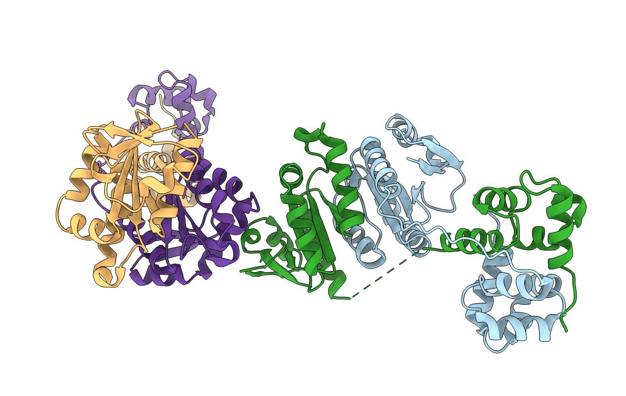
2BHN image
Deposition Date 2005-01-14
Release Date 2005-02-23
Last Version Date 2023-12-13
Entry Detail
PDB ID:
2BHN
Keywords:
Title:
XPF from Aeropyrum pernix
Biological Source:
Source Organism:
Method Details:
Experimental Method:
Resolution:
3.20 Å
R-Value Free:
0.32
R-Value Work:
0.21
R-Value Observed:
0.22
Space Group:
C 1 2 1
Macromolecular Entities
Polymer Type:polypeptide(L)
Molecule:XPF ENDONUCLEASE
Gene (Uniprot):APE_1436.1
Chain IDs:A, B, C, D
Chain Length:214
Number of Molecules:4
Biological Source:Aeropyrum pernix
Ligand Molecules
Primary Citation
Structure of an XPF endonuclease with and without DNA suggests a model for substrate recognition.
EMBO J. 24 895 905 (2005)
PMID: 15719018 DOI: 10.1038/sj.emboj.7600581

Abstact

The XPF/Mus81 structure-specific endonucleases cleave double-stranded DNA (dsDNA) within asymmetric branched DNA substrates and play an essential role in nucleotide excision repair, recombination and genome integrity. We report the structure of an archaeal XPF homodimer alone and bound to dsDNA. Superposition of these structures reveals a large domain movement upon binding DNA, indicating how the (HhH)(2) domain and the nuclease domain are coupled to allow the recognition of double-stranded/single-stranded DNA junctions. We identify two nonequivalent DNA-binding sites and propose a model in which XPF distorts the 3' flap substrate in order to engage both binding sites and promote strand cleavage. The model rationalises published biochemical data and implies a novel role for the ERCC1 subunit of eukaryotic XPF complexes.

Legend

Protein

Chemical

Disease

Primary Citation of related structures