Deposition Date
2005-01-12
Release Date
2005-03-11
Last Version Date
2024-10-09
Entry Detail
PDB ID:
2BHK
Keywords:
Title:
Crystal structure of human growth and differentiation factor 5 (GDF5)
Biological Source:
Source Organism(s):
HOMO SAPIENS (Taxon ID: 9606)
Expression System(s):
Method Details:
Experimental Method:
Resolution:
2.40 Å
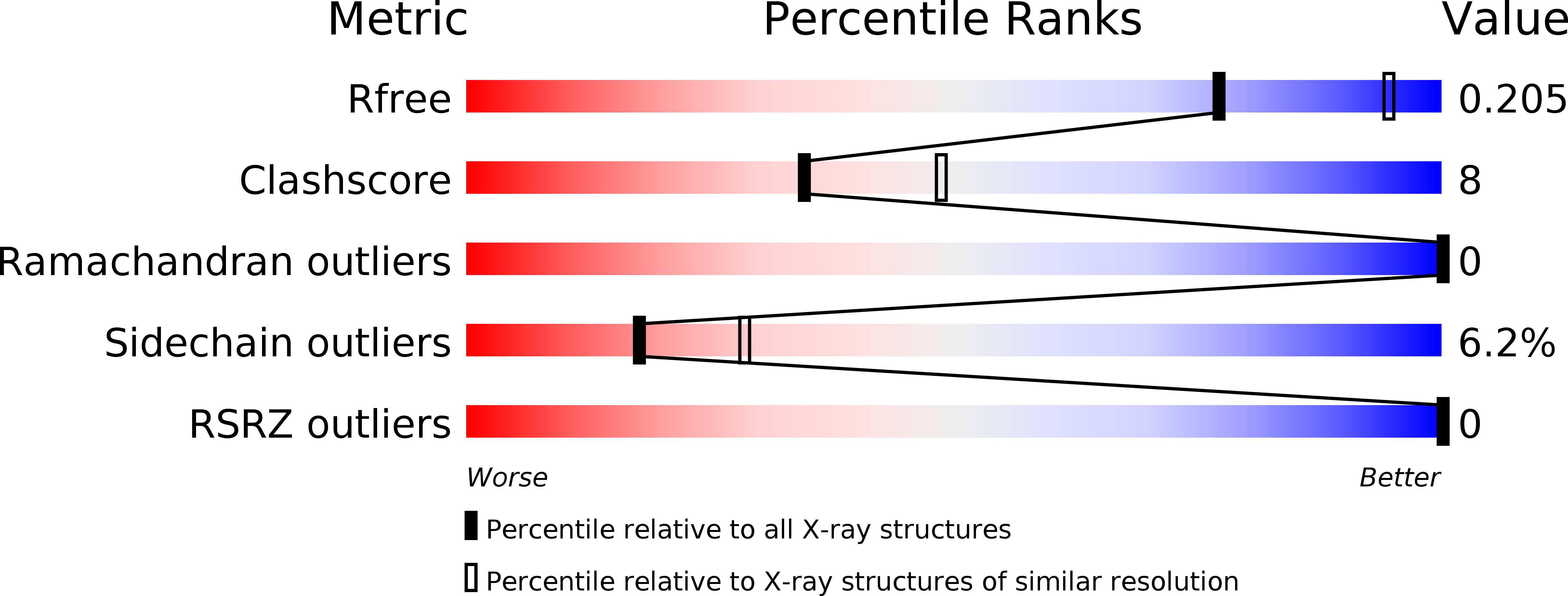
R-Value Free:
0.22
R-Value Work:
0.16
R-Value Observed:
0.16
Space Group:
P 32 2 1