
Deposition Date
2005-01-06
Release Date
2005-05-13
Last Version Date
2023-12-13
Entry Detail
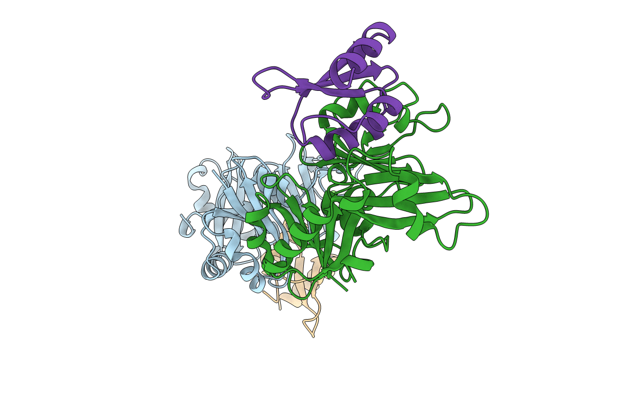
PDB ID:
2BH1
Keywords:
Title:
X-ray structure of the general secretion pathway complex of the N- terminal domain of EpsE and the cytosolic domain of EpsL of Vibrio cholerae
Biological Source:
Source Organism(s):
VIBRIO CHOLERAE (Taxon ID: 666)
Expression System(s):
Method Details:
Experimental Method:
Resolution:
2.40 Å
R-Value Free:
0.25
R-Value Work:
0.19
R-Value Observed:
0.19
Space Group:
P 1 21 1
