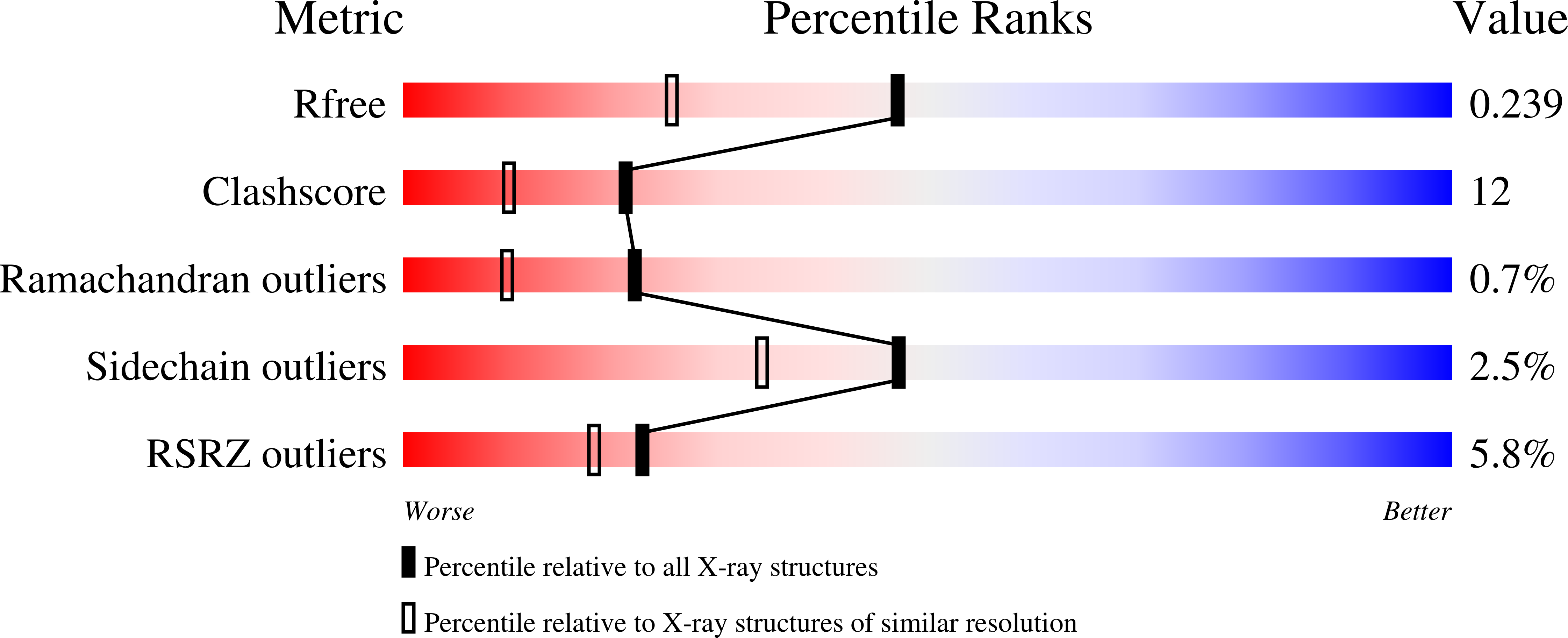
2BDW image
Deposition Date 2005-10-20
Release Date 2005-12-13
Last Version Date 2023-08-23
Entry Detail
PDB ID:
2BDW
Keywords:
Title:
Crystal Structure of the Auto-Inhibited Kinase Domain of Calcium/Calmodulin Activated Kinase II
Biological Source:
Source Organism:
Method Details:
Experimental Method:
Resolution:
1.80 Å
R-Value Free:
0.23
R-Value Work:
0.21
R-Value Observed:
0.21
Space Group:
P 1 21 1
Macromolecular Entities
Structures with similar UniProt ID
Protein Blast
Polymer Type:polypeptide(L)
Molecule:Hypothetical protein K11E8.1d
Mutagens:D135N
Chain IDs:A, B
Chain Length:362
Number of Molecules:2
Biological Source:Caenorhabditis elegans
Primary Citation
Structure of the Autoinhibited Kinase Domain of CaMKII and SAXS Analysis of the Holoenzyme
Cell(Cambridge,Mass.) 123 849 860 (2005)
PMID: 16325579 DOI: 10.1016/j.cell.2005.10.029

Abstact

Ca2+/calmodulin-dependent protein kinase-II (CaMKII) is unique among protein kinases for its dodecameric assembly and its complex response to Ca2+. The crystal structure of the autoinhibited kinase domain of CaMKII, determined at 1.8 A resolution, reveals an unexpected dimeric organization in which the calmodulin-responsive regulatory segments form a coiled-coil strut that blocks peptide and ATP binding to the otherwise intrinsically active kinase domains. A threonine residue in the regulatory segment, which when phosphorylated renders CaMKII calmodulin independent, is held apart from the catalytic sites by the organization of the dimer. This ensures a strict Ca2+ dependence for initial activation. The structure of the kinase dimer, when combined with small-angle X-ray scattering data for the holoenzyme, suggests that inactive CaMKII forms tightly packed autoinhibited assemblies that convert upon activation into clusters of loosely tethered and independent kinase domains.

Legend

Protein

Chemical

Disease

Primary Citation of related structures