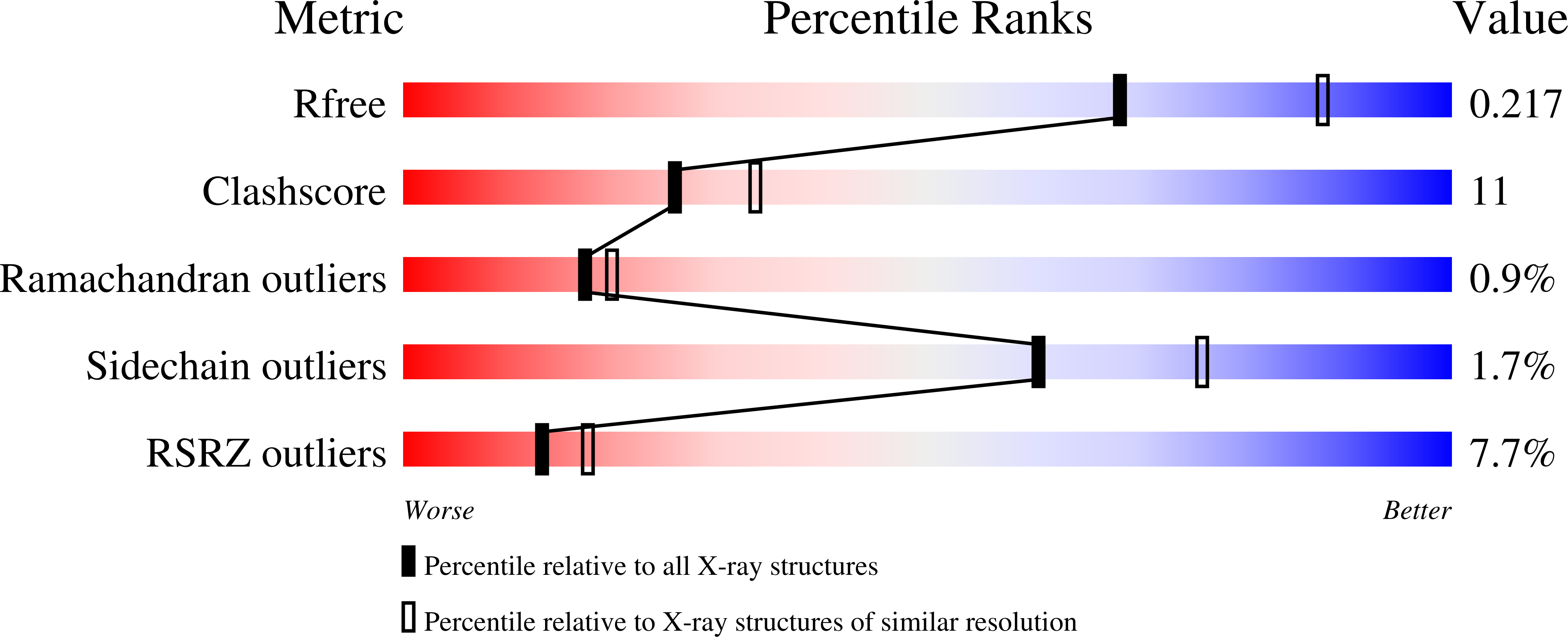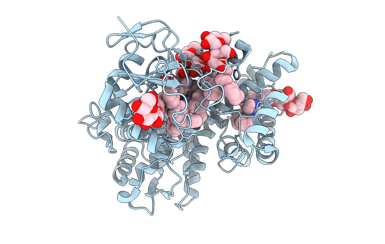
2BDM image
Deposition Date 2005-10-20
Release Date 2005-12-27
Last Version Date 2023-08-23
Entry Detail
PDB ID:
2BDM
Keywords:
Title:
Structure of Cytochrome P450 2B4 with Bound Bifonazole
Biological Source:
Source Organism:
Host Organism:
Method Details:
Experimental Method:
Resolution:
2.30 Å
R-Value Free:
0.21
R-Value Work:
0.19
R-Value Observed:
0.2
Space Group:
P 61 2 2
Macromolecular Entities
Polymer Type:polypeptide(L)
Molecule:Cytochrome P450 2B4
Gene (Uniprot):CYP2B4
Chain IDs:A
Chain Length:476
Number of Molecules:1
Biological Source:Oryctolagus cuniculus
Primary Citation
Structure of microsomal cytochrome P450 2B4 complexed with the antifungal drug bifonazole: insight into P450 conformational plasticity and membrane interaction.
J.Biol.Chem. 281 5973 5981 (2006)
PMID: 16373351 DOI: 10.1074/jbc.M511464200

Abstact

To better understand ligand-induced structural transitions in cytochrome P450 2B4, protein-ligand interactions were investigated using a bulky inhibitor. Bifonazole, a broad spectrum antifungal agent, inhibits monooxygenase activity and induces a type II binding spectrum in 2B4dH(H226Y), a modified enzyme previously crystallized in the presence of 4-(4-chlorophenyl)imidazole (CPI). Isothermal titration calorimetry and tryptophan fluorescence quenching indicate no significant burial of protein apolar surface nor altered accessibility of Trp-121 upon bifonazole binding, in contrast to recent results with CPI. A 2.3 A crystal structure of 2B4-bifonazole reveals a novel open conformation with ligand bound in the active site, which is significantly different from either the U-shaped cleft of ligand-free 2B4 or the small active site pocket of 2B4-CPI. The O-shaped active site cleft of 2B4-bifonazole is widely open in the middle but narrow at the top. A bifonazole molecule occupies the bottom of the active site cleft, where helix I is bent approximately 15 degrees to accommodate the bulky ligand. The structure also defines unanticipated interactions between helix C residues and bifonazole, suggesting an important role of helix C in azole recognition by mammalian P450s. Comparison of the ligand-free 2B4 structure, the 2B4-CPI structure, and the 2B4-bifonazole structure identifies structurally plastic regions that undergo correlated conformational changes in response to ligand binding. The most plastic regions are putative membrane-binding motifs involved in substrate access or substrate binding. The results allow us to model the membrane-associated state of P450 and provide insight into how lipophilic substrates access the buried active site.

Legend

Protein

Chemical

Disease

Primary Citation of related structures