2BBX image
Deposition Date 2005-10-18
Release Date 2006-08-08
Last Version Date 2024-10-30
Entry Detail
PDB ID:
2BBX
Keywords:
Title:
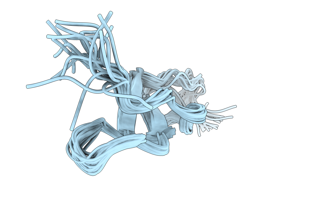
NMR solution structure of the TSR domain of malaria TRAP protein
Biological Source:
Source Organism:
Host Organism:
Method Details:
Experimental Method:
Conformers Calculated:
300
Conformers Submitted:
20
Selection Criteria:
structures with the least restraint violations
Macromolecular Entities
Polymer Type:polypeptide(L)
Molecule:Thrombospondin-related anonymous protein
Gene (Uniprot):TRAP
Chain IDs:A
Chain Length:49
Number of Molecules:1
Biological Source:Plasmodium falciparum
Ligand Molecules
Primary Citation
The layered fold of the TSR domain of P. falciparum TRAP contains a heparin binding site.
Protein Sci. 15 1760 1768 (2006)
PMID: 16815922 DOI: 10.1110/ps.052068506

Abstact

Thrombospondin-related anonymous protein, TRAP, has a critical role in the hepatocyte invasion step of Plasmodium sporozoites, the transmissible form of the parasite causing malaria. The extracellular domains of this sporozoite surface protein interact with hepatocyte surface receptors whereas its intracellular domain acts as a link to the sporozoite actomyosin motor system. Liver heparan sulfate proteoglycans have been identified as potential ligands for TRAP. Proteoglycan binding has been associated with the A- and TSR domains of TRAP. We present the solution NMR structure of the TSR domain of TRAP and a chemical shift mapping study of its heparin binding epitope. The domain has an elongated structure stabilized by an array of tryptophan and arginine residues as well as disulfide bonds. The fold is very similar to those of thrombospondin type-1 (TSP-1) and F-spondin TSRs. The heparin binding site of TRAP-TSR is located in the N-terminal half of the structure, the layered side chains forming an integral part of the site. The smallest heparin fragment capable of binding to TRAP-TSR is a tetrasaccharide.

Legend

Protein

Chemical

Disease

Primary Citation of related structures