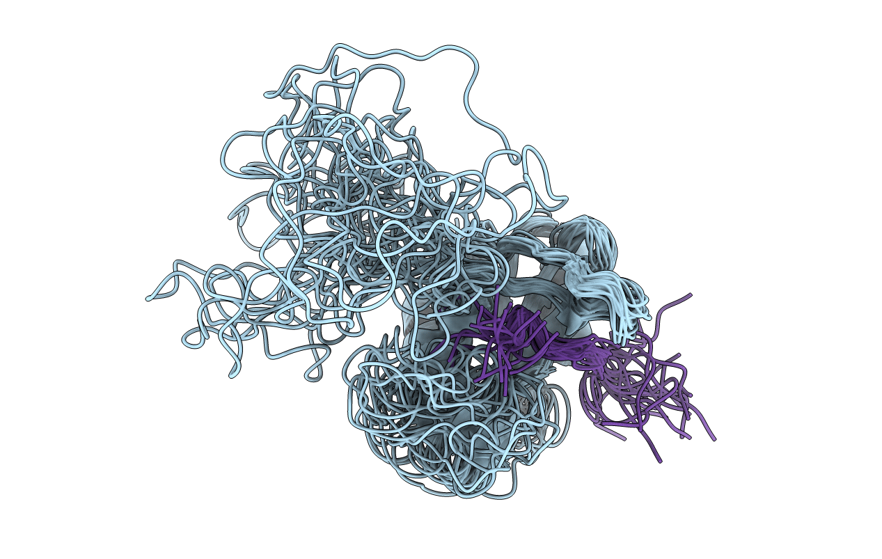
2BBU image
Entry Detail
PDB ID:
2BBU
Title:
solution structure of mouse socs3 in complex with a phosphopeptide from the gp130 receptor
Biological Source:
Source Organism:
Host Organism:
PDB Version:
Deposition Date:
2005-10-17
Release Date:
2006-05-02
Method Details:
Experimental Method:
Conformers Calculated:
200
Conformers Submitted:
20
Selection Criteria:
structures with favorable non-bond energy
Macromolecular Entities
Polymer Type:polypeptide(L)
Description:Suppressor of cytokine signaling 3
Chain IDs:A
Chain Length:164
Number of Molecules:1
Biological Source:Mus musculus
Polymer Type:polypeptide(L)
Description:GP130 PHOSPHOPEPTIDE
Chain IDs:B
Chain Length:15
Number of Molecules:1
Biological Source:
Modified Residue
Compound ID Chain ID Parent Comp ID Details 2D Image
PTR B TYR O-PHOSPHOTYROSINE
Ligand Molecules
Primary Citation
The Structure of SOCS3 Reveals the Basis of the Extended SH2 Domain Function and Identifies an Unstructured Insertion That Regulates Stability
Mol.Cell 22 205 216 (2006)
PMID: 16630890 DOI: 10.1016/j.molcel.2006.03.024

Abstact

SOCS3 is essential for regulating the extent, duration, and specificity of cellular responses to cytokines such as G-CSF and IL-6. Here we describe the solution structure of SOCS3, the first structure determined for any SOCS protein, in complex with a phosphotyrosine-containing peptide from the IL-6 receptor signaling subunit gp130. The structure of the complex shows that seven peptide residues form a predominantly hydrophobic binding motif. Regions outside the SOCS3 SH2 domain are important for ligand binding, in particular, a single 15 residue alpha helix immediately N-terminal to the SH2 domain makes direct contacts with the phosphotyrosine binding loop and, in part, determines its geometry. The SH2 domain itself is remarkable in that it contains a 35 residue unstructured PEST motif insertion that is not required for STAT inhibition. The PEST motif increases SOCS3 turnover and affects its degradation pathway, implying that it has an important regulatory role inside the cell.

Legend

Protein

Chemical

Disease

Primary Citation of related structures