2B9S image
Deposition Date 2005-10-12
Release Date 2006-01-17
Last Version Date 2024-02-14
Entry Detail
PDB ID:
2B9S
Keywords:
Title:
Crystal Structure of heterodimeric L. donovani topoisomerase I-vanadate-DNA complex
Biological Source:
Source Organism:
Method Details:
Experimental Method:
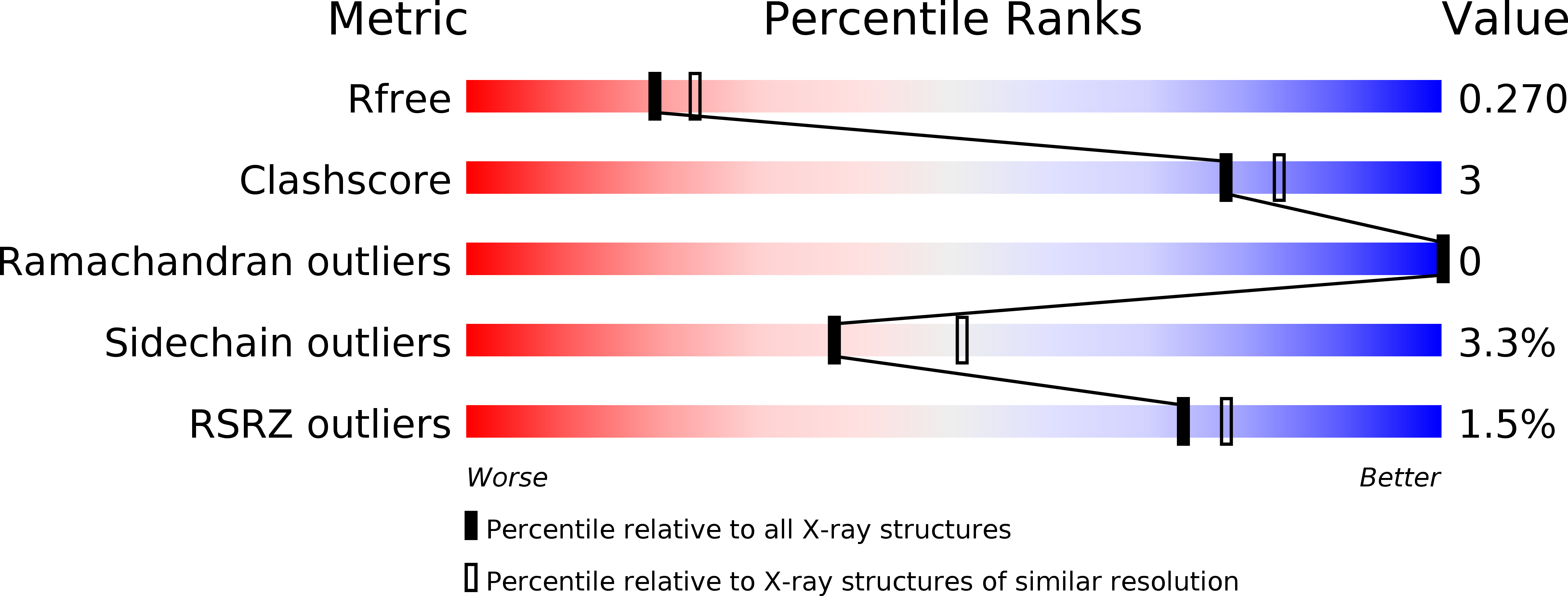
Resolution:
2.27 Å
R-Value Free:
0.27
R-Value Work:
0.23
R-Value Observed:
0.23
Space Group:
P 21 21 21
Macromolecular Entities
Structures with similar UniProt ID
Protein Blast
Polymer Type:polypeptide(L)
Molecule:topoisomerase I-like protein
Chain IDs:D (auth: A)
Chain Length:432
Number of Molecules:1
Biological Source:Leishmania donovani
Structures with similar UniProt ID
Protein Blast
Polymer Type:polypeptide(L)
Molecule:DNA topoisomerase I-like protein
Chain IDs:E (auth: B)
Chain Length:62
Number of Molecules:1
Biological Source:Leishmania donovani
Ligand Molecules
Primary Citation
The Structure of the Transition State of the Heterodimeric Topoisomerase I of Leishmania donovani as a Vanadate Complex with Nicked DNA.
J.Mol.Biol. 357 1202 1210 (2006)
PMID: 16487540 DOI: 10.1016/j.jmb.2006.01.022

Abstact

Type IB topoisomerases are essential enzymes that are responsible for relaxing superhelical tension in DNA by forming a transient covalent nick in one strand of the DNA duplex. Topoisomerase I is a target for anti-cancer drugs such as camptothecin, and these drugs also target the topoisomerases I in pathogenic trypanosomes including Leishmania species and Trypanosoma brucei. Most eukaryotic enzymes, including human topoisomerase I, are monomeric. However, for Leishmania donovani, the DNA-binding activity and the majority of residues involved in catalysis are located in a large subunit, designated TOP1L, whereas the catalytic tyrosine residue responsible for covalent attachment to DNA is located in a smaller subunit, called TOP1S. Here, we present the 2.27A crystal structure of an active truncated L.donovani TOP1L/TOP1S heterodimer bound to nicked double-stranded DNA captured as a vanadate complex. The vanadate forms covalent linkages between the catalytic tyrosine residue of the small subunit and the nicked ends of the scissile DNA strand, mimicking the previously unseen transition state of the topoisomerase I catalytic cycle. This structure fills a critical gap in the existing ensemble of topoisomerase I structures and provides crucial insights into the catalytic mechanism.

Legend

Protein

Chemical

Disease

Primary Citation of related structures