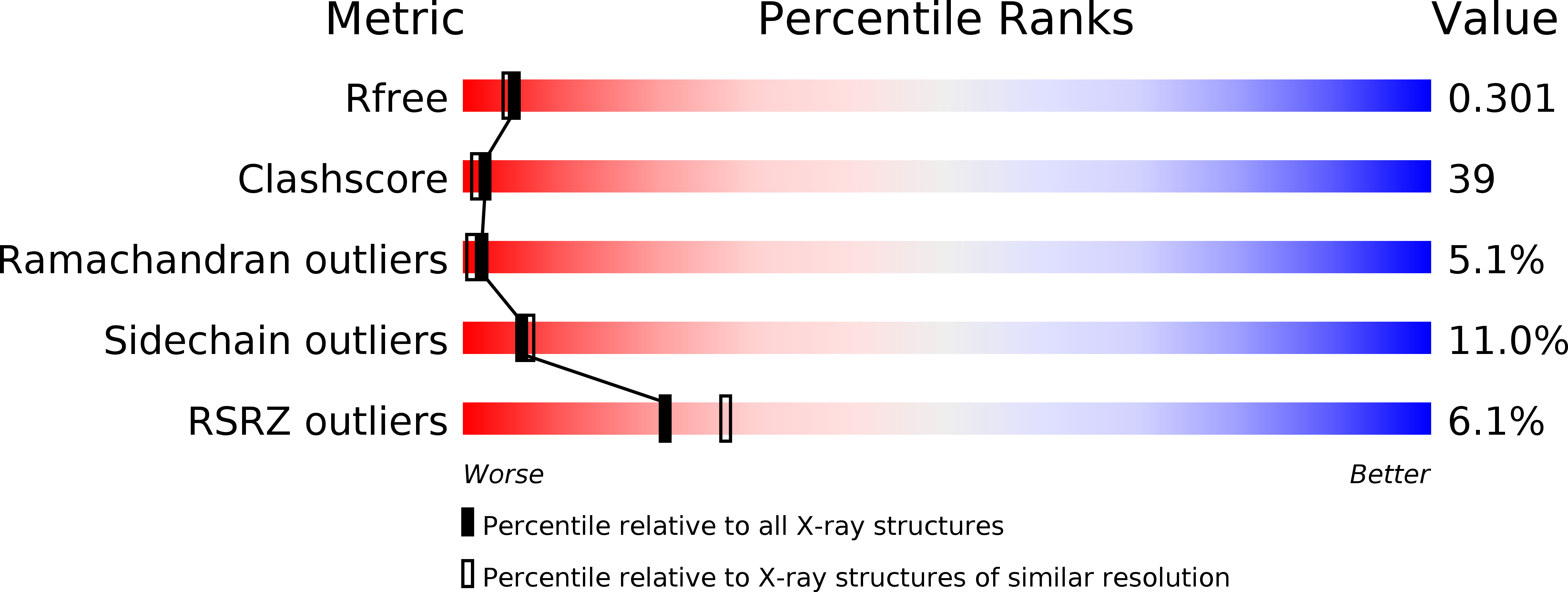
2B9C image
Deposition Date 2005-10-11
Release Date 2006-01-03
Last Version Date 2024-10-30
Entry Detail
PDB ID:
2B9C
Title:
Structure of tropomyosin's mid-region: bending and binding sites for actin
Biological Source:
Source Organism:
Host Organism:
Method Details:
Experimental Method:
Resolution:
2.30 Å
R-Value Free:
0.29
R-Value Work:
0.25
Space Group:
P 65
Macromolecular Entities
Structures with similar UniProt ID
Protein Blast
Polymer Type:polypeptide(L)
Molecule:striated-muscle alpha tropomyosin
Gene (Uniprot):Tpm1
Chain IDs:A, B
Chain Length:147
Number of Molecules:2
Biological Source:Rattus norvegicus
Primary Citation
Structure of the mid-region of tropomyosin: Bending and binding sites for actin.
Proc.Natl.Acad.Sci.Usa 102 18878 18883 (2005)
PMID: 16365313 DOI: 10.1073/pnas.0509269102

Abstact

Tropomyosin is a two-chain alpha-helical coiled coil whose periodic interactions with the F-actin helix are critical for thin filament stabilization and the regulation of muscle contraction. Here we deduce the mechanical and chemical basis of these interactions from the 2.3-A-resolution crystal structure of the middle three of tropomyosin's seven periods. Geometrically specific bends of the coiled coil, produced by clusters of core alanines, and variable bends about gaps in the core, produced by isolated alanines, occur along the molecule. The crystal packing is notable in signifying that the functionally important fifth period includes an especially favorable protein-binding site, comprising an unusual apolar patch on the surface together with surrounding charged residues. Based on these and other results, we have constructed a specific model of the thin filament, with the N-terminal halves of each period (i.e., the so-called "alpha zones") of tropomyosin axially aligned with subdomain 3 of each monomer in F-actin.

Legend

Protein

Chemical

Disease

Primary Citation of related structures