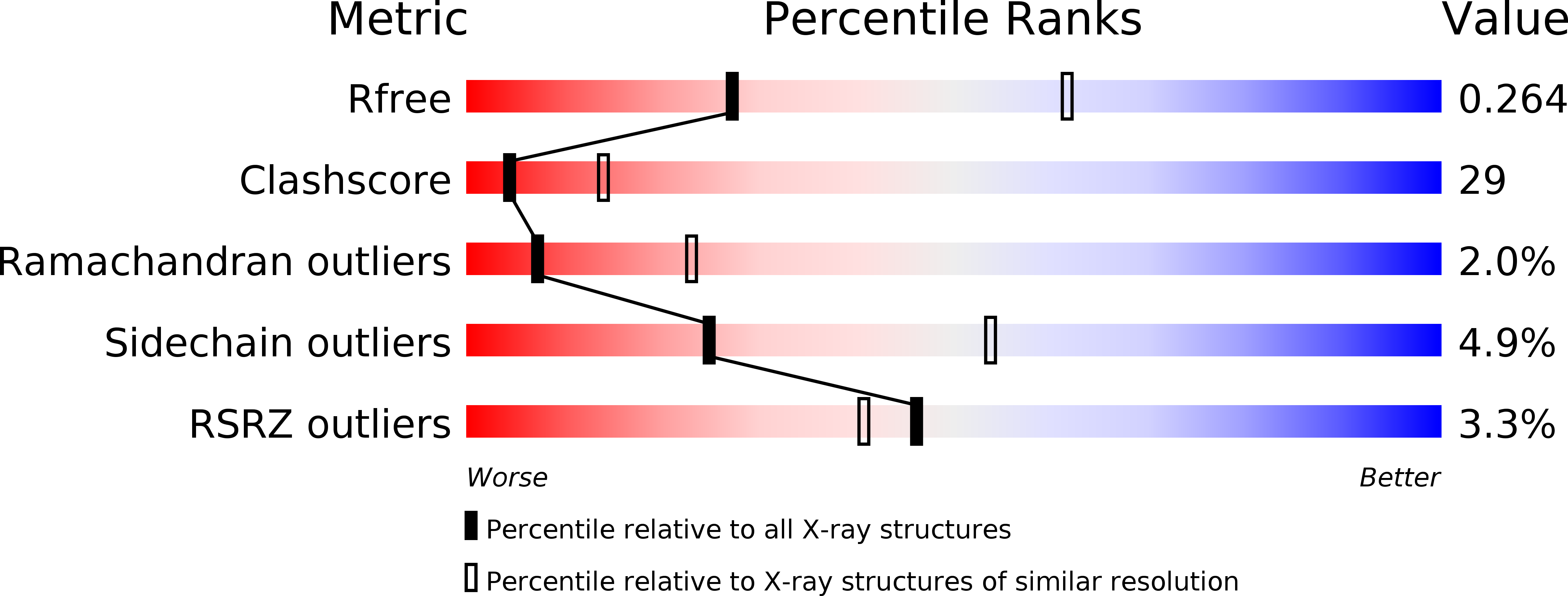
2B9B image
Deposition Date 2005-10-11
Release Date 2006-01-24
Last Version Date 2024-11-20
Entry Detail
PDB ID:
2B9B
Keywords:
Title:
Structure of the Parainfluenza Virus 5 F Protein in its Metastable, Pre-fusion Conformation
Biological Source:
Source Organism:
Simian virus 5 (Taxon ID: 11207)
Host Organism:
Method Details:
Experimental Method:
Resolution:
2.85 Å
R-Value Free:
0.25
R-Value Work:
0.22
R-Value Observed:
0.22
Space Group:
C 2 2 21
Macromolecular Entities
Structures with similar UniProt ID
Protein Blast
Polymer Type:polypeptide(L)
Molecule:Fusion glycoprotein F0
Gene (Uniprot):F
Chain IDs:A, B, C
Chain Length:497
Number of Molecules:3
Biological Source:Simian virus 5
Modified Residue
Compound ID Chain ID Parent Comp ID Details 2D Image
ASN A ASN GLYCOSYLATION SITE
Ligand Molecules
Primary Citation
Structure of the parainfluenza virus 5 F protein in its metastable, prefusion conformation
Nature 439 38 44 (2006)
PMID: 16397490 DOI: 10.1038/nature04322

Abstact

Enveloped viruses have evolved complex glycoprotein machinery that drives the fusion of viral and cellular membranes, permitting entry of the viral genome into the cell. For the paramyxoviruses, the fusion (F) protein catalyses this membrane merger and entry step, and it has been postulated that the F protein undergoes complex refolding during this process. Here we report the crystal structure of the parainfluenza virus 5 F protein in its prefusion conformation, stabilized by the addition of a carboxy-terminal trimerization domain. The structure of the F protein shows that there are profound conformational differences between the pre- and postfusion states, involving transformations in secondary and tertiary structure. The positions and structural transitions of key parts of the fusion machinery, including the hydrophobic fusion peptide and two helical heptad repeat regions, clarify the mechanism of membrane fusion mediated by the F protein.

Legend

Protein

Chemical

Disease

Primary Citation of related structures