2B92 image
Deposition Date 2005-10-10
Release Date 2006-03-07
Last Version Date 2023-08-23
Entry Detail
PDB ID:
2B92
Title:
Crystal-structure of the N-terminal Large GTPase Domain of human Guanylate Binding protein 1 (hGBP1) in complex with GDP/AlF3
Biological Source:
Source Organism:
Homo sapiens (Taxon ID: 9606)
Host Organism:
Method Details:
Experimental Method:
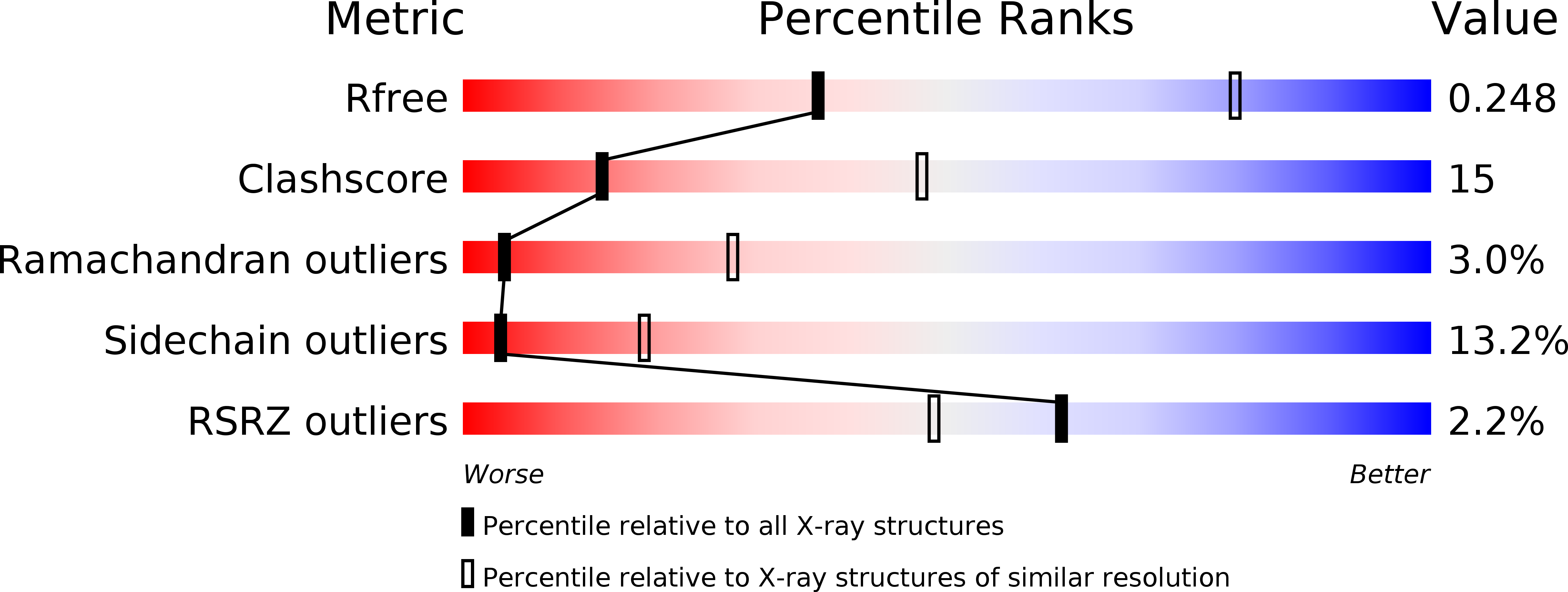
Resolution:
3.20 Å
R-Value Free:
0.25
R-Value Work:
0.20
R-Value Observed:
0.20
Space Group:
P 21 21 21
Macromolecular Entities
Polymer Type:polypeptide(L)
Molecule:Interferon-induced guanylate-binding protein 1
Gene (Uniprot):GBP1
Chain IDs:A, B
Chain Length:328
Number of Molecules:2
Biological Source:Homo sapiens
Primary Citation
How guanylate-binding proteins achieve assembly-stimulated processive cleavage of GTP to GMP.
Nature 440 101 104 (2006)
PMID: 16511497 DOI: 10.1038/nature04510

Abstact

Interferons are immunomodulatory cytokines that mediate anti-pathogenic and anti-proliferative effects in cells. Interferon-gamma-inducible human guanylate binding protein 1 (hGBP1) belongs to the family of dynamin-related large GTP-binding proteins, which share biochemical properties not found in other families of GTP-binding proteins such as nucleotide-dependent oligomerization and fast cooperative GTPase activity. hGBP1 has an additional property by which it hydrolyses GTP to GMP in two consecutive cleavage reactions. Here we show that the isolated amino-terminal G domain of hGBP1 retains the main enzymatic properties of the full-length protein and can cleave GDP directly. Crystal structures of the N-terminal G domain trapped at successive steps along the reaction pathway and biochemical data reveal the molecular basis for nucleotide-dependent homodimerization and cleavage of GTP. Similar to effector binding in other GTP-binding proteins, homodimerization is regulated by structural changes in the switch regions. Homodimerization generates a conformation in which an arginine finger and a serine are oriented for efficient catalysis. Positioning of the substrate for the second hydrolysis step is achieved by a change in nucleotide conformation at the ribose that keeps the guanine base interactions intact and positions the beta-phosphates in the gamma-phosphate-binding site.

Legend

Protein

Chemical

Disease

Primary Citation of related structures