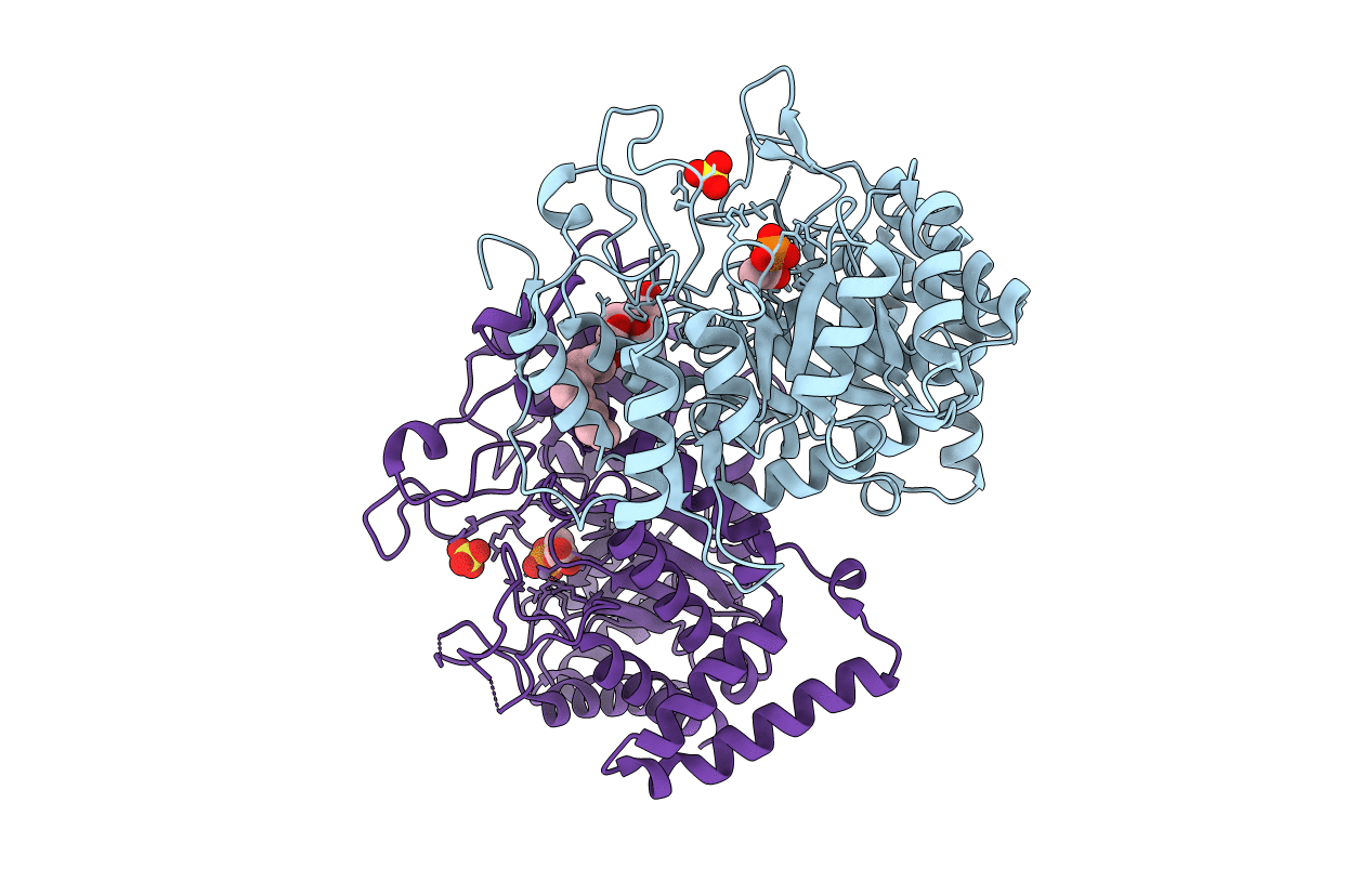
2B7O image
Entry Detail
PDB ID:
2B7O
Keywords:
Title:
The Structure of 3-Deoxy-D-Arabino-Heptulosonate 7-Phosphate Synthase from Mycobacterium tuberculosis
Biological Source:
Host Organism:
PDB Version:
Deposition Date:
2005-10-05
Release Date:
2005-10-18
Method Details:
Experimental Method:
Resolution:
2.30 Å
R-Value Free:
0.22
R-Value Work:
0.18
R-Value Observed:
0.18
Space Group:
P 32 2 1
Macromolecular Entities
Polymer Type:polypeptide(L)
Description:3-deoxy-D-arabino-heptulosonate 7-phosphate synthase AroG
Chain IDs:A, B
Chain Length:464
Number of Molecules:2
Biological Source:Mycobacterium tuberculosis
Modified Residue
Compound ID Chain ID Parent Comp ID Details 2D Image
MSE A MET SELENOMETHIONINE
Primary Citation
The structure of 3-deoxy-d-arabino-heptulosonate 7-phosphate synthase from Mycobacterium tuberculosis reveals a common catalytic scaffold and ancestry for type I and type II enzymes
J.Mol.Biol. 354 927 939 (2005)
PMID: 16288916 DOI: 10.1016/j.jmb.2005.09.093

Abstact

The shikimate pathway, responsible for the biosynthesis of aromatic compounds, is essential for the growth of Mycobacterium tuberculosis and is a potential target for the design of new anti-tuberculosis drugs. The first step of this pathway is catalyzed by 3-deoxy-d-arabino-heptulosonate 7-phosphate synthase (DAH7PS). The DAH7PSs have been classified into two apparently unrelated types and, whereas structural data have been obtained for the type I DAH7PSs, no structural information is available for their type II counterparts. The type II DAH7PS from M.tuberculosis has been expressed in Escherichia coli, purified, functionally characterized and crystallized. It is found to be metal ion-dependent and subject to feedback inhibition by phenylalanine, tryptophan, tyrosine and chorismate, with a significant synergistic effect when tryptophan is used in combination with phenylalanine. The crystal structure of M.tuberculosis DAH7PS has been determined by single-wavelength anomalous diffraction and refined at 2.3A in complex with substrate phosphoenolpyruvate and Mn(2+). The structure reveals a tightly associated dimer of (beta/alpha)(8) TIM barrels. The monomer fold, the arrangement of key residues in the active site, and the binding modes of PEP and Mn(2+), all match those of the type I enzymes, and indicate a common ancestry for the type I and type II DAH7PSs, despite their minimal sequence identity. In contrast, the structural elements that decorate the core (beta/alpha)(8) fold differ from those in the type I enzymes, consistent with their different regulatory and oligomeric properties.

Legend

Protein

Chemical

Disease

Primary Citation of related structures