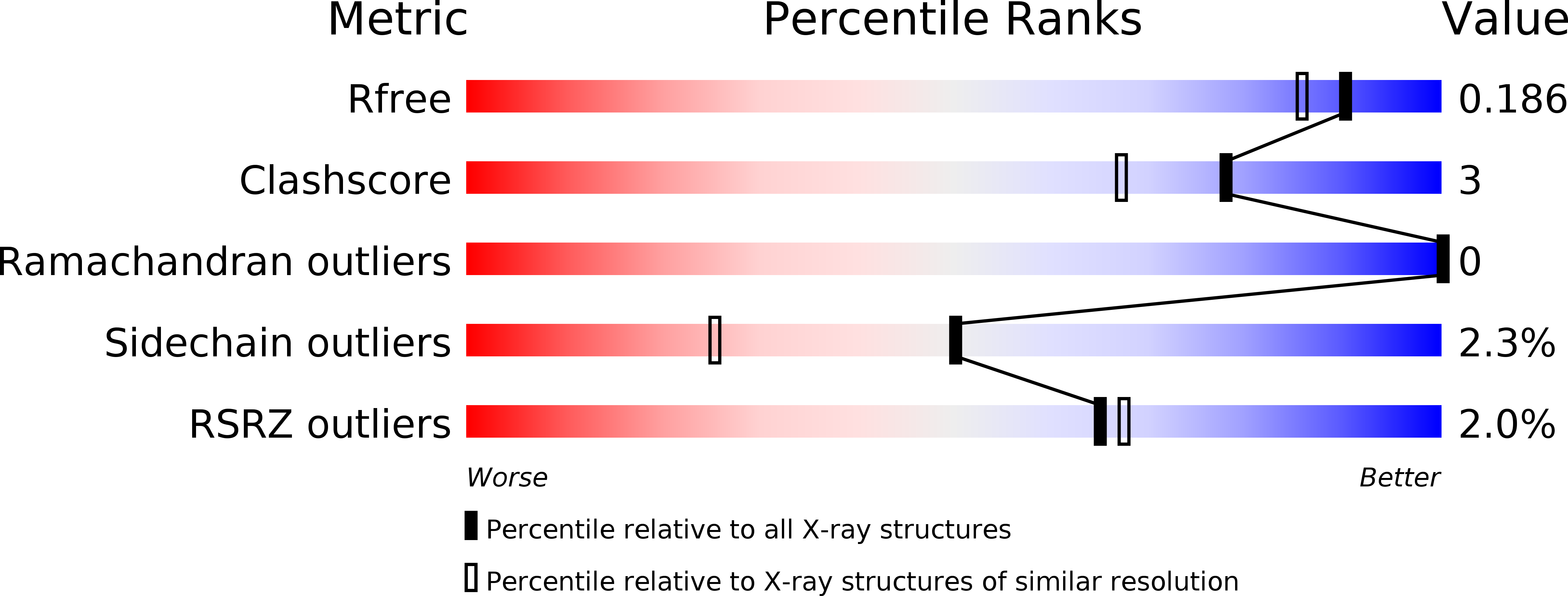
2B61 image
Deposition Date 2005-09-29
Release Date 2005-11-15
Last Version Date 2024-11-13
Entry Detail
PDB ID:
2B61
Keywords:
Title:
Crystal Structure of Homoserine Transacetylase
Biological Source:
Source Organism:
Host Organism:
Method Details:
Experimental Method:
Resolution:
1.65 Å
R-Value Free:
0.18
R-Value Work:
0.16
R-Value Observed:
0.16
Space Group:
P 31 2 1
Macromolecular Entities
Polymer Type:polypeptide(L)
Molecule:Homoserine O-acetyltransferase
Gene (Uniprot):metXA
Chain IDs:A
Chain Length:377
Number of Molecules:1
Biological Source:Haemophilus influenzae
Modified Residue
Compound ID Chain ID Parent Comp ID Details 2D Image
MSE A MET SELENOMETHIONINE
Primary Citation
Crystal Structure of Homoserine Transacetylase from Haemophilus influenzae Reveals a New Family of alpha/beta-Hydrolases
Biochemistry 44 15768 15773 (2005)
PMID: 16313180 DOI: 10.1021/bi051951y

Abstact

Homoserine transacetylase catalyzes one of the required steps in the biosynthesis of methionine in fungi and several bacteria. We have determined the crystal structure of homoserine transacetylase from Haemophilus influenzae to a resolution of 1.65 A. The structure identifies this enzyme to be a member of the alpha/beta-hydrolase structural superfamily. The active site of the enzyme is located near the end of a deep tunnel formed by the juxtaposition of two domains and incorporates a catalytic triad involving Ser143, His337, and Asp304. A structural basis is given for the observed double displacement kinetic mechanism of homoserine transacetylase. Furthermore, the properties of the tunnel provide a rationale for how homoserine transacetylase catalyzes a transferase reaction vs hydrolysis, despite extensive similarity in active site architecture to hydrolytic enzymes.

Legend

Protein

Chemical

Disease

Primary Citation of related structures